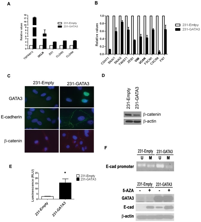
Figure 1. GATA3 over-expression in MB-231 cells promotes EMT and the re-expression of E-cadherin.
(A) Q-RT-PCR demonstrates increased expression of epithelial markers in 231-GATA3 cells compared to 231-Empty cells. We observed increased TSPAN13, OCLN, ZO1, CLDN3 and CLDN4 in 231-GATA3 vs. 231-Empty cells. Samples were normalized to cyclophilin B. (B) Q-RT-PCR showing that the relative expression of mesenchymal and metastatic markers is reduced in 231-GATA3 cells compared to 231-Empty cells. We observed reduced CDH1, SNAI1, SNAI2, TWIST1, ZEB1, VIM, VCAN, FSCN1, CXCR4 and FN1 in 231-GATA3 vs. 231-Empty cells. Samples were normalized to cyclophilin B. (C) Immunofluorescence of 231-Empty and 231-GATA3 cells for GATA3, E-cadherin and ß-catenin. GATA3 over-expression in MB-231 results in re-expression of E-cadherin associated with the cell membrane and re-distribution of ß-catenin. (D) Western blot showing reduced expression of ß-catenin in 231-GATA3 cells compared to 231-Empty cells. ß-actin was used as loading control. (E) Luciferase activity showing increased activity of the E-cadherin reporter construct in 231-GATA3 cells vs. 231-Empty cells. (F) top panel: PCR using primers that detect only unmethylated or methylated DNA at the E-cadherin promoter of 231-Empty and 231-GATA3 cells. DNA underwent bisulfate treatment prior to the PCR reaction. 231-GATA3 cells show reduced methylated and increased unmethylated DNA in the E-cadherin promoter region compared to 231-Empty cells. Lower panel: western blot of 231-Empty and 231-GATA3 cells for E-cadherin. Cells were treated with 5-AZA for 4 days prior to lysate collection. Treatment of 231-Empty and 231-GATA3 cells with 5-AZA increased E-cadherin expression.

