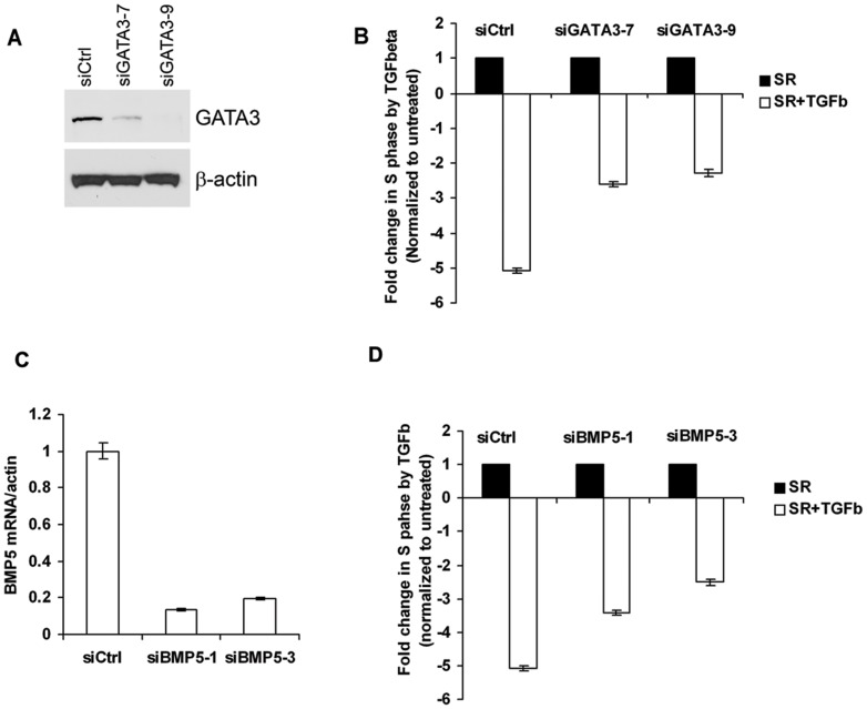
Figure 8. GATA3 and BMP5 knock down in MIII mammary epithelial cells decrease growth suppressive effect of TGFß.
(A) Western blot analysis of GATA3 in MIII cells transfected with control or GATA-3 siRNA. (B) Flow cytometric analysis of MIII cells after transfection with control or GATA3 siRNA and TGFßtreatment. (C) Quantitative RT-PCR analysis of BMP5 expression after transfection with control or BMP5 siRNA. (D) Flow cytometric analysis of MIII cells after transfection with control or BMP5 siRNA and TGFßtreatment. More cells were in proliferative state (S-phase) after knockdown of GATA-3 (B) or BMP5 (D) and subsequent TGFß treatment than control siRNA-transfected cells treated with TGFß All values were normalized to that of the TGFß untreated cells.

