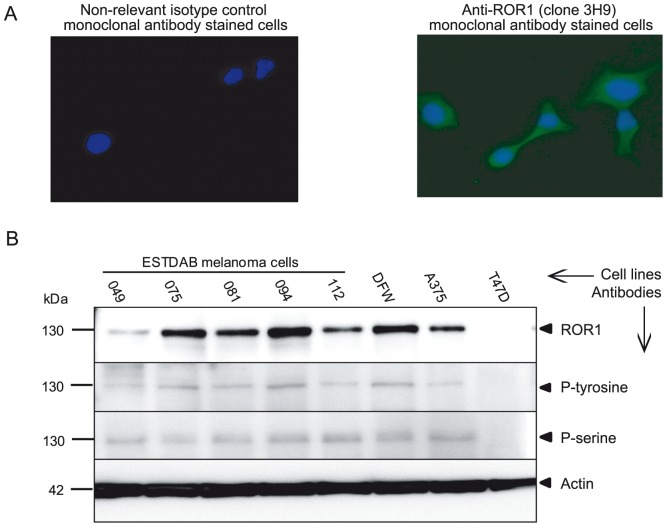
Figure 1. Protein expression of the receptor tyrosine kinase ROR1 in melanoma cell lines.
Representative experiment (IF) showing the expression of ROR1 on the ESTDAB112 cell line using the anti-ROR1 (clone 3H9) mAb (40×). Nuclei were counterstained with DAPI (blue). A non-relevant isotype control mAb (mouse IgG1 isotype) was used as a negative control (A). Western blot analysis of ROR1 protein expression and phosphorylation in melanoma cells detected by a goat anti-ROR1 antibody, anti-p-tyrosine (PY99) and anti-p-serine (clone 4A4) mAbs (B). ROR1 protein was shown to be phosphorylated in all cell lines using immunoprecipitation of ROR1. A 130 kDa band corresponding to the fully glycosylated/phosphorylated ROR1 was observed. The T47D cell line was used as a ROR1 negative control [16].

