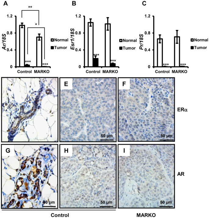
Figure 5. Steroid receptor expression is reduced in MMTV-NeuNT tumors.
At the time of sacrifice, tumors were harvested and non-tumor bearing mammary glands were dissociated from the fat pad. RNA was prepared as described in the methods section. Levels of Ar (A), Esr1 (B), Pr (C) and 18S were analyzed by quantitative RT-PCR (Control normal = 26, Control tumor = 9, and MARKO normal = 15 and MARKO tumor = 7). Expression of each receptor was normalized to 18S. Receptors in tumors for MARKO and Control mice and normal MARKO glands were normalized to expression in non-tumor bearing mammary glands from the Control group. *denotes P<0.05, **P<0.01 and ***P<0.005. Immunohistochemical detection of ERα in the normal mammary gland (D) of Control mice and tumors from Control (E) and MARKO (F) mice. Immunohistochemical detection of AR in the normal mammary gland of Control mice (G) and tumors from Control (H) and MARKO (I) mice. Scale bar = 50 µm.

