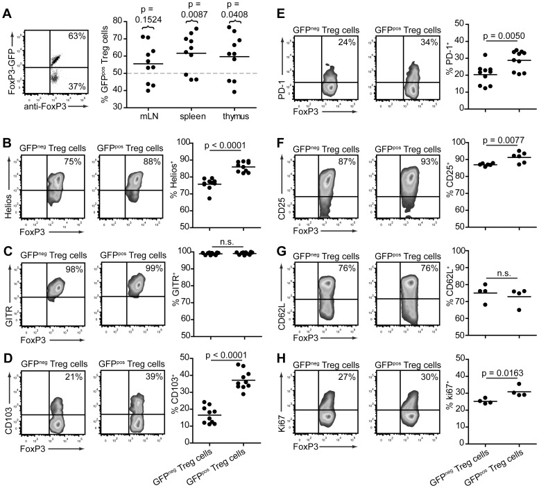
Figure 2. GFPpos Treg cells display a potentially more suppressive phenotype in Tg4 FoxP3wt/gfp females.
A. Representative dot plot (splenocytes, left) and distribution graph (right) showing the frequency of GFP expression in CD4+FoxP3+ cells from the thymus, spleen and mLN of Tg4 FoxP3wt/gfp females aged 5–8 weeks. Horizontal dotted line represents the predicted frequency. P values show significance of actual vs predicted value. 2-tailed, paired student's t test. n = 10 each. B-H. Representative, smoothed, cytometry plots and distribution graphs showing the frequency of marker expression on GFPpos and GFPneg splenic CD4+FoxP3+ Treg cells in Tg4 FoxP3wt/gfp females aged 5–10 weeks. P values represent significance determined by 2-tailed, unpaired student's t test. n.s. = not significant. B. Helios, n = 10. C. GITR, n = 10. D. CD103, n = 10. E. PD-1, n = 10. F. CD25, n = 6. G. CD62L, n = 4. H. Ki67, n = 4.

