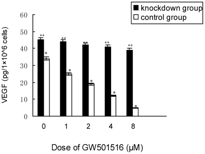
Figure 6. KM12C cells secreted less VEGF after PPAR δ activation.

The KM12C cells were treated with serial concentrations of GW501516 or vehicle, and the cell-free supernatants were collected for VEGF quantification by ELISA. Treated by GW501516, the control cells showed a dose-dependent decrease of VEGF secretion (*P = 0.018), while the PPAR δ-silenced cells had no significant change all along (**P = 0.83; ANOVA).
