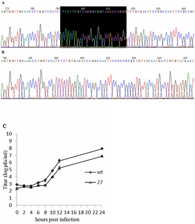
Figure 3. Sequence analysis and growth kinetics measurements of MHV-A59 and MHV-nsp1-27D.
(A) WT MHV-A59 sequence. (B) MHV-nsp1-27D sequence, with deletion of nts 780–807 of nsp1. (C) Comparison of MHV-A59 and MHV-nsp1-27D growth. 17Cl-1 cells were infected with MHV-A59 or MHV-nsp1-27D at an MOI of 1 pfu/cell. Samples of culture medium were obtained at 0, 2, 4, 6, 8, 10, 12 and 24 h p.i., and viral titers were determined by plaque assay. Each time point indicates the mean titer and standard deviation obtained from a triplicate series of infections.

