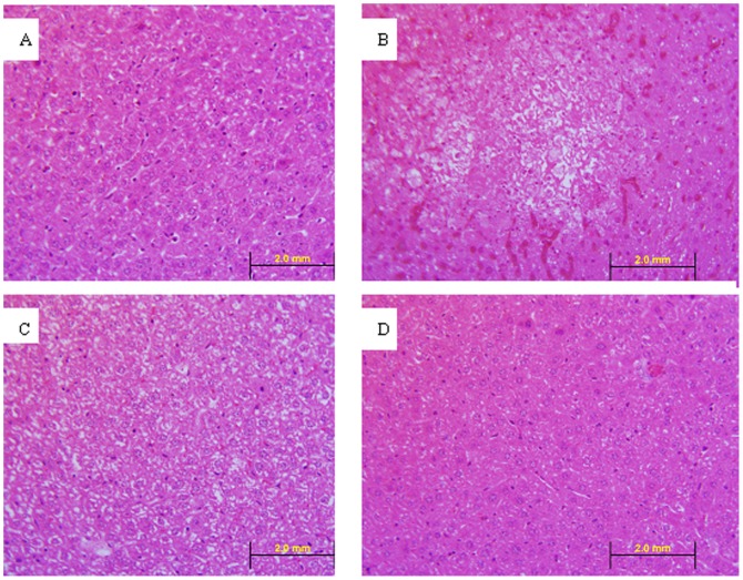
Figure 6. Hematoxylin and eosin staining of liver sections (40×).
A: Liver section from a normal mice. B: Liver section from a mouse of the MHV-WT group, on day 5 (intra-abdominal infection, 5×10³ pfu/animal). C: Liver section from a mouse of the MHV-nsp1-27D group, on day 8 (intra-abdominal infection, 5×10³ pfu/animal). D: Liver section from a mouse of the MHV-nsp1-27D group on day 15 (intra-abdominal infection, 5×10³ pfu/animal). The liver of the MHV-WT infected mice showed obvious lesions on day 5. The hepatocytes in the center of the lesion were fibroblast-like and without nuclei. In MHV-nsp1-27D infected mice, the hepatocytes showed unclear borderlines and increased gaps on day 8, which was restored to a normal state on day 15.

