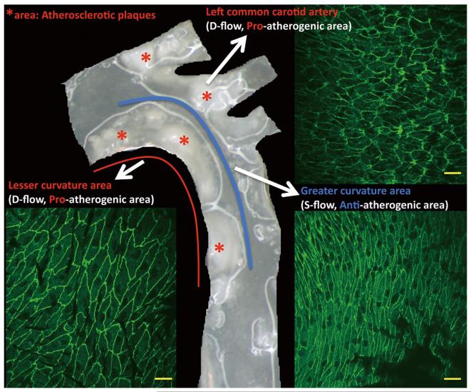
Figure 1.
Morphology of endothelial cells and atherosclerotic plaques in the arch region of the mouse aorta. The greater curvature of the aortic arch and the straight region of the aorta indicated by the blue line are exposed to s-flow and are protected from atherosclerosis. Regions of curvature indicated by the red line and side branches are exposed to d-flow and are atheroprone areas. The aorta was prepared from a LDLR−/− mouse fed with high cholesterol diet for 16 weeks; red stars indicate atherosclerotic plaques. En face endothelial cell morphology illustrated by anti-VE-cadherin staining was prepared from the aortic arch of a wild-type mouse. Scale bars, 20μm. d-flow, disturbed flow; s-flow, steady laminar flow; LDLR−/−; low-density lipoprotein receptor knockout.

