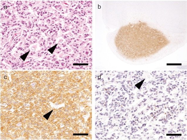
Fig 3.
H&E and immunohistochemical staining of Case 3 (SD rat).a: Neoplastic cells proliferate in the piriform lobe, and reactive astrocytes are observed (arrow). H&E stain. Bar=50 μm. b: Iba-1 shows strong immunoreactivityin the region of neoplastic cell infiltration. Bar=1 mm. c: Neoplastic cells are strongly positive for Iba-1, but reactive astrocytes are negative. Bar=50 μm. d: Neoplastic cells are sporadically positive for CD68, but the reactive astrocytes exhibit negative immunoreactivity. Bar=50 μm.

