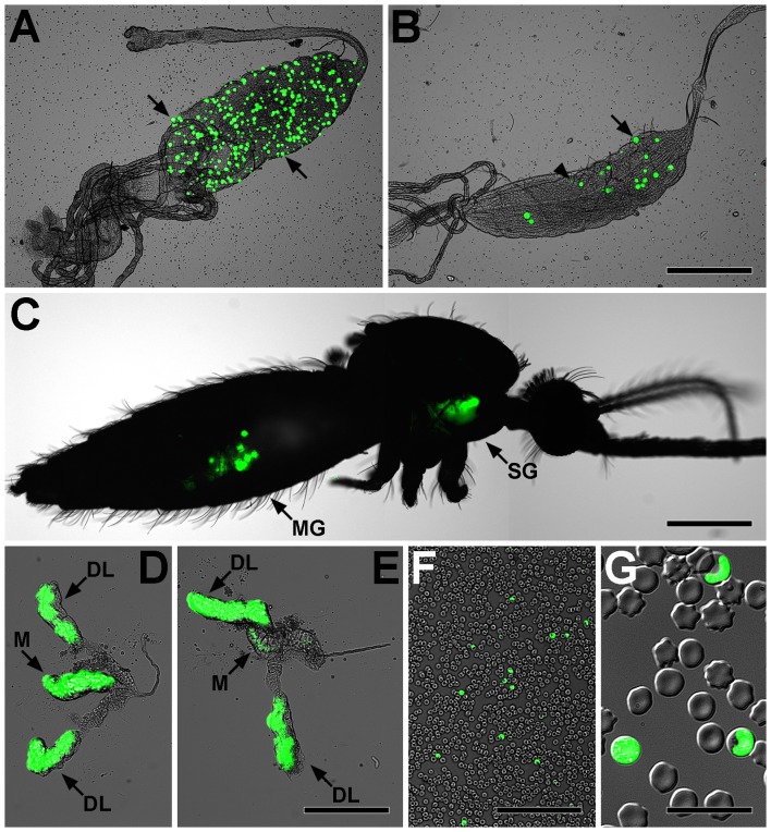
Figure 1. Light and GFP epi-fluorescence microscopy show fluorescent Plasmodium berghei parasites developing in Anopheles funestus mosquitoes and murine erythrocytes.
A. A. funestus midgut with greater than 300 P. berghei oocysts (e.g., arrows) at 7 days post-infection. B. A. funestus midgut showing normal oocysts (e.g., arrow) and oocysts that have recently undergone rupture (e.g., arrowhead) at 20 days post-infection. C. Imaging of fluorescent parasites through the cuticle of a live A. funestus showing parasite development in the midgut (MG) and salivary glands (SG). Note that tissues presented in panels B, D, and E originated from this mosquito. D–E. A. funestus salivary glands showing that sporozoites preferentially invade the median (M) and distal lateral (DL) lobes. F–G. Blood smear from a mouse exposed to P. berghei via mosquito bite showing infected erythrocytes, indicating that A. funestus salivary gland sporozoites are infective to the vertebrate host and that the parasite can complete its life cycle inside the insect vector. Bars: A–C = 500 µm; D–E = 200 µm; F = 100 µm; G = 20 µm.

