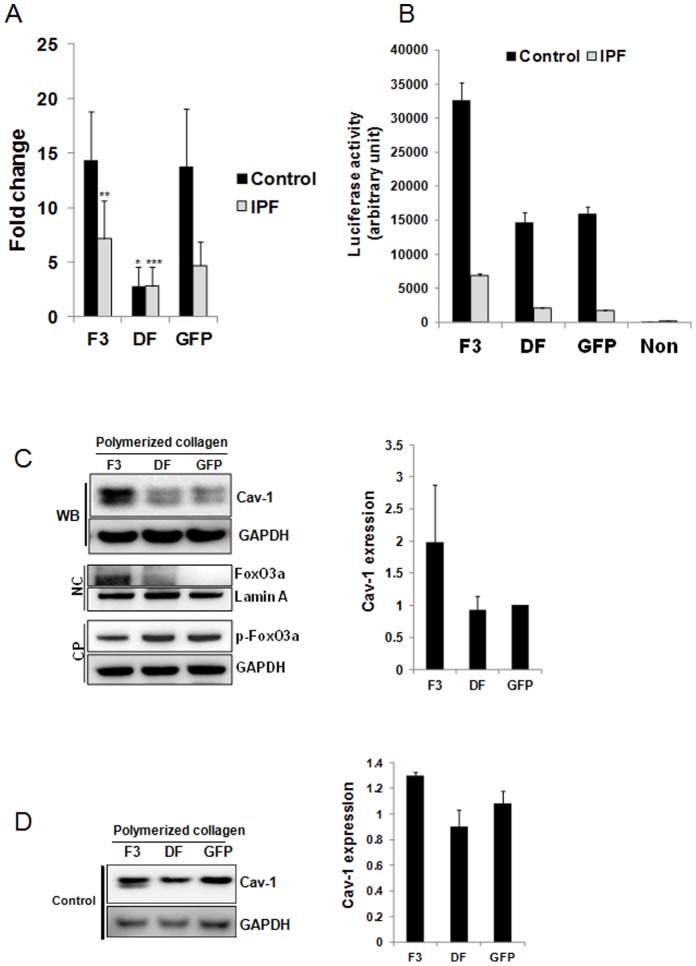
Figure 3. FoxO3a deficiency suppresses cav-1 expression in IPF fibroblast on collagen matrix.
A. IPF and control fibroblasts infected with adenovirus expressing wild type FoxO3a (F3), dominant negative FoxO3a (DF) or empty vector (GFP) were cultured on polymerized collagen for 24 h in serum free medium. After total RNA was isolated, quantitative RT-PCR was carried out with cav-1 primers as described in the Materials and Methods. Shown is quantitative RT-PCR for cav-1 mRNA levels normalized to 18S rRNA in IPF and control fibroblasts. *p = 0.03 versus GFP in control fibroblasts, **p = 0.02 versus GFP in IPF fibroblasts. ***p = 0.016 versus GFP in IPF fibroblast. The assay was obtained from triplicate from 2 control and 1 IPF fibroblasts. B. IPF and control fibroblasts expressing wild type FoxO3a (F3), mutant FoxO3a (DF) or empty vector (GFP) were transfected with a luciferase construct containing consensus Forkhead binding sites (FHRE-Luc). Cells were attached to polymerized collagen in serum free medium for 24 h and luciferase activity was measured as described in the Materials and Methods. Non: cells without transfection of FHRE-Luc construct. The assay was obtained from triplicate. C. Left upper panel, shown is representative Western blot analysis (WB) of cav-1 protein expression in IPF fibroblasts expressing wild type (F3), dominant negative FoxO3a (DF) or GFP control and cultured on polymerized collagen for 24 h. Left middle panel, nuclear fraction was isolated from IPF fibroblasts cultured on collagen as described in the Materials and Methods and unphosphorylated FoxO3a protein level was measured. NC : nuclear fraction. Lamin A was used as a loading control. Left lower panel : cytoplasmic fraction was isolated from IPF fibroblasts cultured on collagen and phosphorylated FoxO3a (p-FoxO3a) was measured. CP : cytoplasmic fraction. GAPDH is shown as a loading control. Right panel, densitometric analysis of cav-1/GAPDH expression ratio. D. Left panel, control fibroblasts expressing wild type (F3), dominant negative FoxO3a (DF) or GFP control and cultured on polymerized collagen for 24 h and cav-1 protein expression was measured. GAPDH is shown as loading control. Right panel, densitometric analysis of cav-1/GAPDH expression ratio. All blots represent 3 independent assays.

