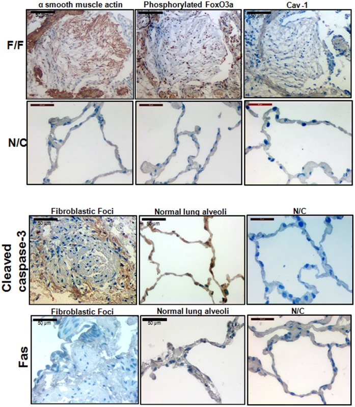
Figure 7. Fibroblasts within the fibroblastic foci of IPF patient lung specimens express inactive FoxO3a and α-smooth muscle actin but not cav-1 and cleaved caspase-3.
Upper upper panel, frozen serial sections of IPF patient lung specimens (F/F: fibroblastic foci) were immunostained with anti-α smooth muscle actin antibody (upper left), phosphorylated FoxO3a (inactive FoxO3a, upper middle), or anti-cav-1 (upper right), and IHC was carried out as described in the Materials and Methods (n = 3). Upper lower panel, shown are IHC images obtained from control tissue without primary antibodies as negative controls (N/C). Lower upper panel. IHC analysis of IPF (left) and control (middle) lung tissue was performed using anti-cleaved caspase-3 antibodies. N/C : IHC images obtained from control tissue without cleaved caspase-3 antibody as a negative control. Lower lower panel. Fas expression was measured from frozen sections of IPF (left) and control (middle) lung specimens. N/C : IHC images obtained from control tissue without Fas antibody as a negative control. Note that cav-1, cleaved caspase-3 and Fas expression were absent or very low in cells within the fibroblastic foci.

