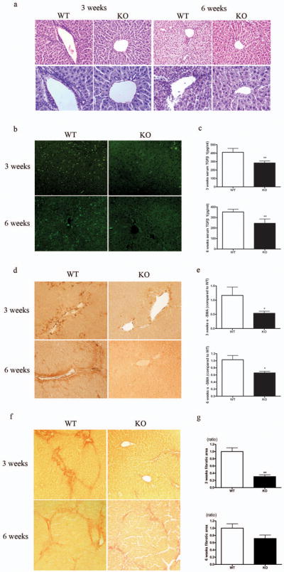
Figure 3.

SRC-3−/− mice were protected against chronic hepatic fibrosis. (a) Representative H&E stained liver sections from SRC-3+/+ and SRC-3−/− mice treated chronically with CCl4 for 3 and 6 weeks, respectively. Inflammatory infiltration was significantly attenuated in SRC-3−/− mice compared to SRC-3+/+ mice (upper panels, ×100 magnification; lower panels, ×200 magnification). (b) Immunofluerence staining for CD8 positive cells in livers from SRC-3+/+ and SRC-3−/− mice treated chronically with CCl4 for 3 and 6 weeks, respectively (×100 magnification). (c) Quantification of serum TGFβ1 level by ELISA in SRC-3+/+ and SRC-3−/− mice treated chronically with CCl4 for 3 and 6 weeks, respectively. (d) Immunohistochemical analysis of α-SMA expression in livers from SRC-3+/+ and SRC-3−/− mice treated chronically with CCl4 for 3 and 6 weeks, respectively (×100 magnification). (e) Expression of α-SMA mRNA levels was analyzed in livers from SRC-3+/+ and SRC-3−/− mice treated chronically with CCl4 for 3 and 6 weeks, respectively. All data were normalized against house keeping gene β-actin and expressed as mean fold change in SRC-3−/− samples relative to SRC-3+/+. (f) Representative Sirius Red stained liver sections from SRC-3+/+ and SRC-3−/− mice treated chronically with CCl4 for 3 and 6 weeks, respectively. Livers from SRC-3−/− mice had less collagen deposition compared to SRC-3+/+ mice (×100 magnification). (g) Digital quantification of Sirius Red staining on liver sections from SRC-3+/+ and SRC-3−/− mice treated chronically with CCl4 for 3 and 6 weeks, respectively. Data were expressed as mean fold change in SRC-3−/− samples relative to SRC-3+/+. All data are mean ± standard error. *P<0.05, **P<0.01, WT vs KO.
