
Figure 6.
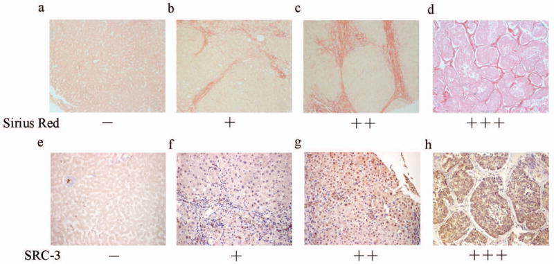
Positive correlation between SRC-3 and fibrosis degrees in human hepatic diseases. (a-d) Fibrosis extents in liver samples from patients with viral hepatitis or hepatic cirrhosis were examined by Sirius Red staining and representative images were shown for grading and grouping. (a) Negative staining of hepatitis. (b)+ of hepatitis. (c) ++ of hepatitis. (d) strong staining (+++) of hepatic cirrhosis (×100 Magnification). (e-h) SRC-3 nuclues immunoreactivity in the same samples shown in Figure 6a-d. They are also representative images for the grading and grouping of SRC-3 immunoreactivity. (e) Negative staining of hepatitis. (f)+ of hepatitis. (g) ++ of hepatitis. (h) strong staining (+++) of hepatic cirrhosis (×200 Magnification).