Figure 3.
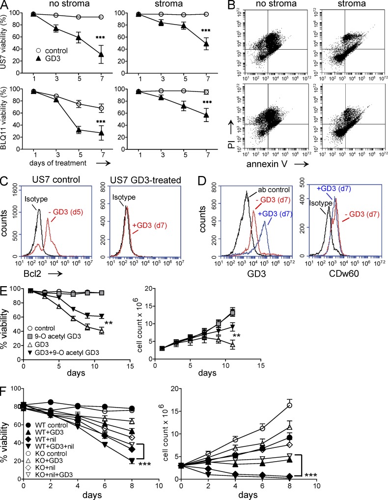
Exogenously added GD3 causes apoptosis of human and mouse pre-B ALL cells. (A) Viability of human pre-B ALLs in the absence (left) or presence (right) of irradiated OP9 stroma after treatment with 20 µM GD3. Experiments were performed independently three times. Control versus GD3: ***, P < 0.001 day 7. (B) Annexin V/PI staining of GD3 treated cells from A at day 7. (C) Bcl-2 expression before and after 7 d of exposure to 20 µM GD3 in US7 cells in the presence of stroma. The experiment was independently replicated once. (D) Expression of total GD3 and 9-O-acetylated GD3 on day 7 of 20-µM GD3 treatment in the presence of stroma. Samples are as indicated. The experiment was performed four times. (E) Viability (left) and viable cell numbers (right) of US7 cells treated with 50 µM GD3, 100 µM 9-O-Ac–GD3, or a combination of both. GD3 and 9-O-Ac–GD3 were added every alternate day. Untreated cells served as control. **, P < 0.01 (viability and cell counts day 12) for GD3- compared with GD3 + 9-O-Ac–GD3–treated cells. (F) Viability (left) and viable cell numbers (right) of BCR/ABL-transduced WT and KO cells not treated or treated with 50 µM GD3, 12 nM nilotinib, or both. Experiments in E and F were performed twice. Error bars show the standard deviation of triplicate samples. ***, P < 0.001 (viability and cell count d8) for WT + GD3 + nil compared with KO + GD3 + nil.

