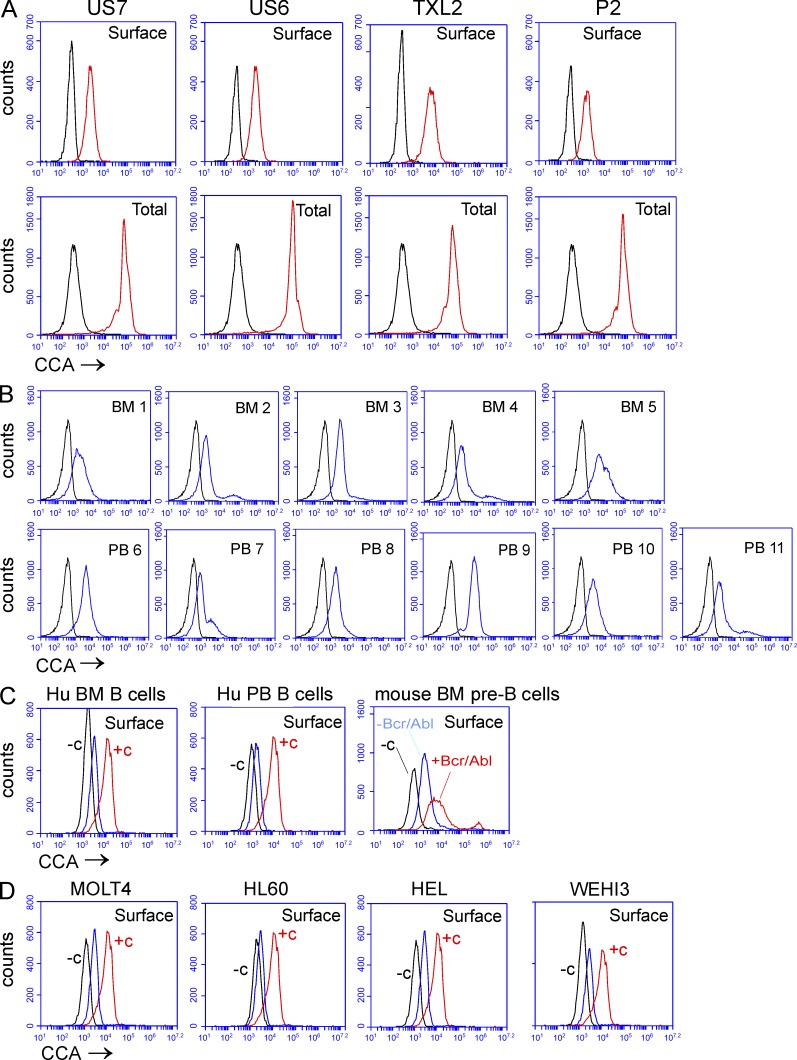
Figure 4.
Human ALL cells express O-acetylated Neu5Ac detected by FITC–CCA lectin. FACS analysis showing cell surface binding of FITC–CCA lectin. (A) human ALL cells co-cultured with mouse stroma (black, control signal; red, CCA lectin signal). (B) CD19+, CD10+ pre-B cells from BM and PB of ALL patients (black, control; blue, CCA lectin). (C) Normal human BM B cells (left), normal human PB B cells (middle), or WT pre-B cells (red) with or (blue) without BCR/ABL transduction. (D) Non-ALL leukemia cells. In C and D, −c (black) indicates controls without CCA lectin, and +c indicates US7 staining (red) as positive reference sample; CCA lectin binding is shown in blue.

