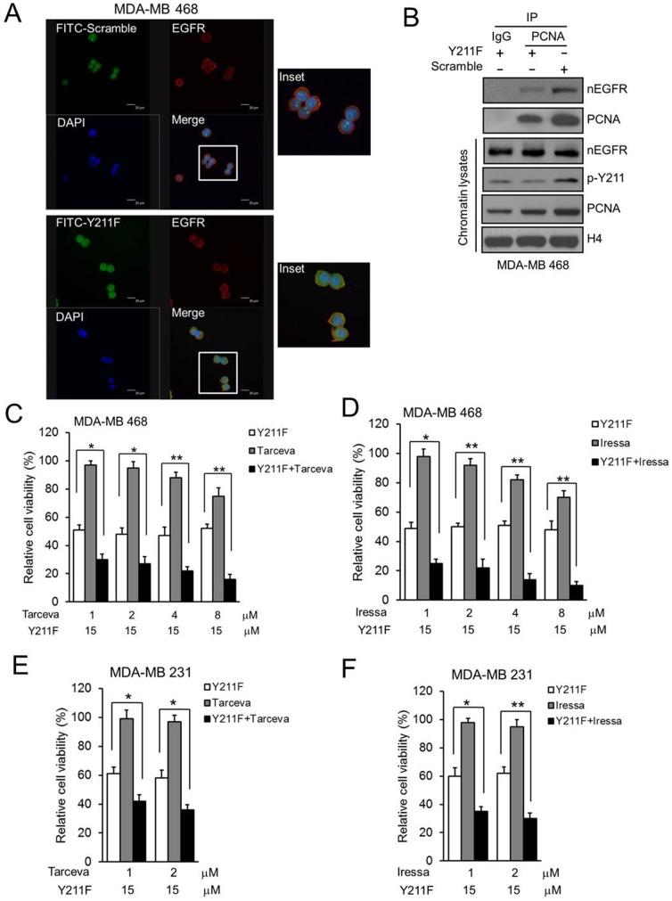
Figure 2. The effect of CPPP on PCNA Y211 phosphorylation and EGFR TKIs.
(A) MDA-MB 468 cells were treated with 30 µM FITC-labeled scrambled (FITC-Scrambled; top) or Y211F CPPP (FITC-Y211F; bottom) for 12 h. Subsequently, cells were fixed, permeabilized, and stained with anti-EGFR antibody or DAPI. The fluorescent images were observed under a confocal microscopy. The FITC-labeled peptide (green), EGFR (red), and nucleus (blue) are shown. (B) MDA-MB 468 cells were treated with 30 µM scrambled (Scramble) or Y211F CPPP (Y211F) for 24 h. The chromatin lysates were extracted and IP with IgG or anti-PCNA antibody and separated by SDS-PAGE followed by IB for nEGFR, Y211-phosphorylated PCNA (p-Y211) and PNCA in MDA-MB 468 cells. (C–F) Both MDA-MB 468 or MDA-MB 231 cells were treated with 15 µM Y211F CPPP or 1∼8 µM EGFR TKI (Tarceva or Iressa) alone, or Y211F CPPP and TKI combined for 24 h. The relative cell viability after each treatment was then determined.

