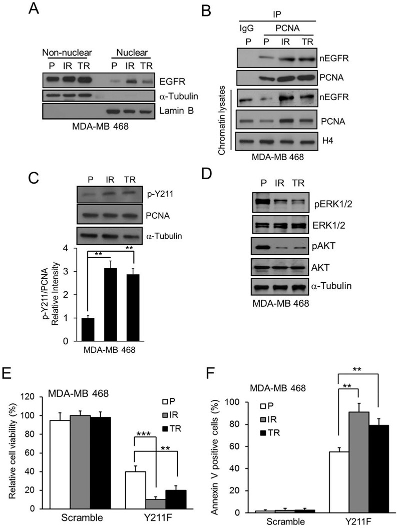
Figure 3. The cytotoxic effect of CPPP in TKI-resistant TNBC cells.
(A) Cell lysate from non-nuclear and nuclear fractions were isolated from parental (P), Iressa-resistant (IR) or Tarceva-resistant (TR) MDA-MB 468 cells. The protein level of EGFR in each fraction was examined by IB. α-tubulin and lamin B were used as the markers of non-nuclear and nuclear fraction, respectively. (B) The association between nEGFR and PCNA in chromatin lysate from P-, IR- and TR-TNBC cells were verified by IP with IgG or anti-PCNA antibody. (C) The amount of p-Y211 on PCNA in each TNBC line was examined. A comparison of the relative intensity p-Y211 in IR- or TR-TNBC cells to the parental TNBCs is shown at the bottom. (D) Total cell lysate from P-, IR- and TR-TNBC cells were isolated. The protein level of indicated proteins were examined by IB. (E–F) The P-, IR-, or TR-TNBC cells were treated with 15 µM Y211F CPPP for 24 h and the relative cell viability (E) and Annexin V-positive cells (F) were determined. The bars indicate mean ± S.D. **, p<0.01; ***, p<0.001 by t-test.

