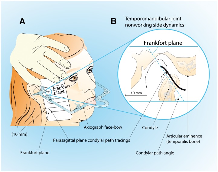
Figure 1. Axiography procedure: condylar path tracings.
A, the kinematic face-bow attached with silicone putty to mandibular teeth through an occlusal rim; lateral condylar path drawn on the surface of the recording card. B, parasagittal plane of lateral condylar path tracings and their angle with respect to the tragus-infraorbital Frankfort plane.

