Fig. 2.

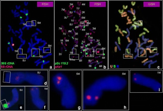
a–i Mitotic metaphase cell division of Ae. ovata × S. cereale (plant 3/4) analysed using: a FISH pattern showing the location of 5S rDNA (red) and 35S rDNA (green), followed by b FISH pattern showing the location of pSc119.2 (green) and pAs1 (red) repetitive clones and c genomic in situ hybridization (GISH) with total genomic DNA of Ae. umbellulata (red), Ae. comosa (green) used as probes and S. cereale (blue) used as blocking DNA. d Chromosome 5U with 35S rDNA deletion, e chromosome 5U, f, i chromosome 1M, g, h chromosome 5M
