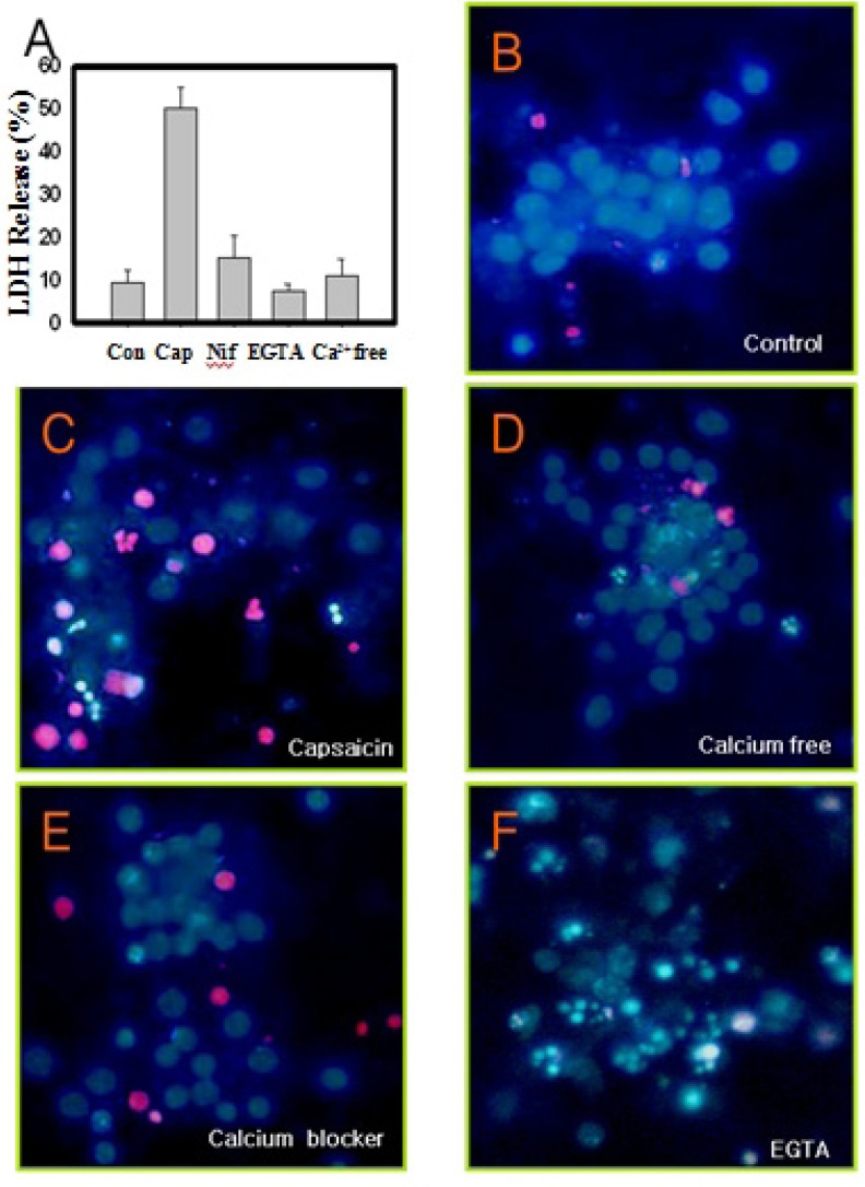
Fig. 3.
Measurement of cell death and fluorescence micrographs (Hoechst-PI stain) of primary cortex neurons pretreated with calcium blocker, followed by 10 µM capsaicin treatment. (A) LDH assay, (B) control, (C) capsaicin only, (D) calcium free, (E) 1 µM nifedipine, (F) 50 uM EGTA. Capsaicin-induced cell death were attenuated when a nifedipine, calcium channel blocker, and calcium free media were applied.

