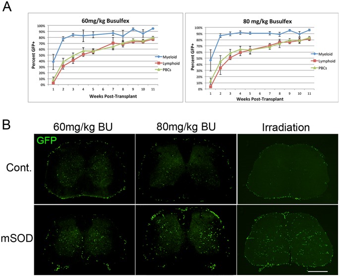
Figure 2. PBC reconstitution by donor cells and distribution of GFP+ cells in lumbar spinal cord.
(A) Donor PBC reconstitution in mice treated with 60 mg/kg BU and 80 mg/kg BU was assessed weekly for 11 weeks post-transplant using flow cytometry. Levels of PBC chimerism achieved were similar between 60 mg/kg BU and 80 mg/kg BU treatment groups. Reconstitution of myelomonocytic cells was rapid in comparison to lymphoid populations, owing to the shorter half-life of circulating myelomonocytic cells and the minimal immunosuppressive effects of BU. Error bars represent standard deviation. (B) Distribution of GFP+ cells in control and mSOD lumbar spinal cord sections 11–14 weeks after transplantation following BU treatment or myeloablative irradiation. GFP+ cells accumulated in the grey and white matter of lumbar spinal cord sections, especially in the surrounding leptomeninges 11–14 weeks after transplantation following BU treatment or irradiation. Significantly greater numbers of BMDCs accumulated in mSOD lumbar spinal cords compared to age-matched controls using BU (60 or 80 mg/kg) or irradiative myelosuppression (p<0.05). Scale bar = 500 µm.

