
Figure 1.
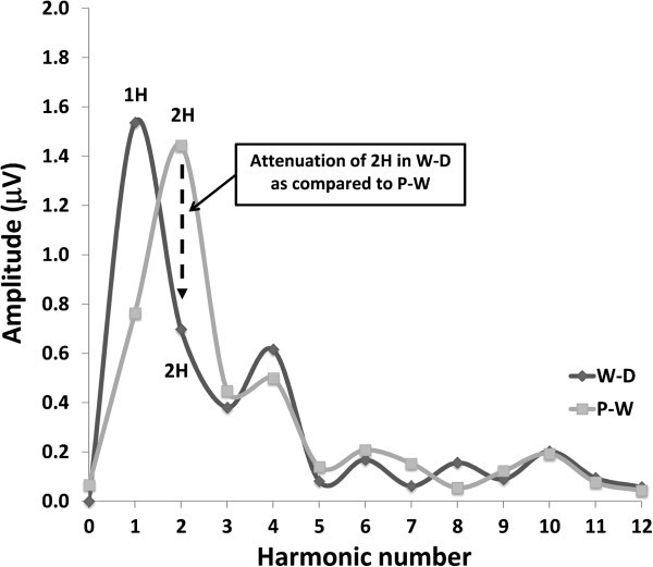
Illustrative traces of visual evoked potentials obtained by checkerboard (left column), windmill-dartboard (middle column) and partial-windmill (right column) stimulation in a healthy volunteer (upper row) and a migraine without aura patient between attacks (lower row).