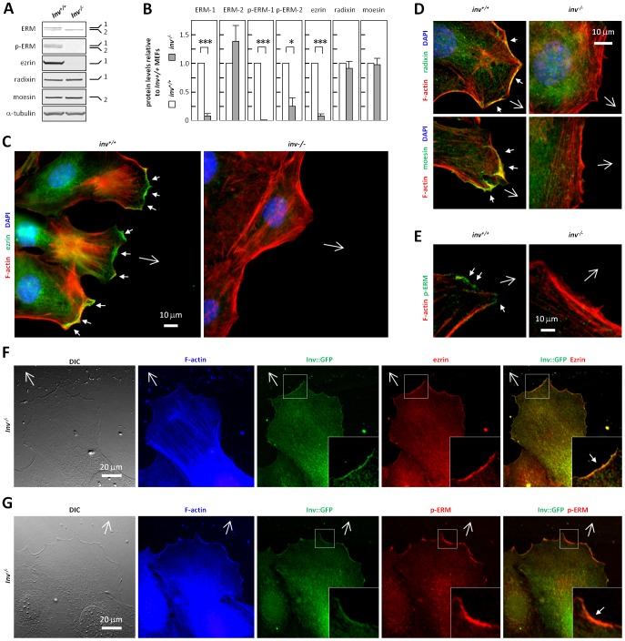
Figure 6. Inversin affects expression, regulation and localization of ERM proteins.
(A) WB analysis of growth arrested inv+/+ and inv−/− MEFs with antibodies against total ezrin/radixin/moesin (ERM), phosphorylated ERM (T567 of ezrin, T564 of Radixin, T558 of moesin, p-ERM), ezrin, radixin and moesin, and α-tubulin as control, with indications of the 80 (1) and 75 (2) kDa bands. (B) Quantification of WB from (A); Histograms represent mean ± S.E.M. (n≥3). (C,E) IFM analysis of growth arrested inv+/+ and inv−/− MEFs in wound healing assays, with phalloidin staining of the actin cytoskeleton (F-actin, red) and nuclei with DAPI (blue). Open arrows indicate direction of migration, and arrowhead indicate leading edge staining of ezrin (C, green), Radixin (D; upper panel, green), moesin (D; lower panel, green), and p-ERM (E, green). (F,G) DIC and IFM analysis on lamellipodium formation and localization of ERM (F, red) and p-ERM (G, red) to the leading edge of migrating cells in wound healing assays in Inv-GFP (green) transfected inv−/− MEFs. The actin cytoskeleton is stained with phalloidin (F-actin, blue). Open arrows indicate direction of migration.

