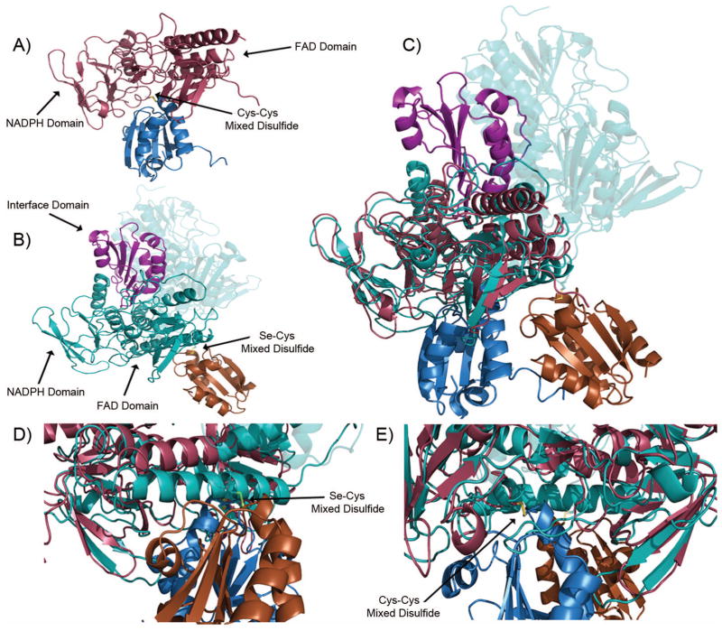
Figure 5.
Comparison of the M. tuberculosis. and human Trx-TrxR complex. (a) M. tuberculosis. Trx (blue)-TrxR (red) mixed disulfide. (b) Human Trx (orange)-TrxR (teal) mixed disulfide. Note the carboxy-terminus of the dimer pair is highlighted in violet and forms the mixed disulfide. (c) Overlay of the M. tuberculosis. and human thioredoxin systems. (d) Close up of M. tuberculosis thioredoxin complex disulfide from Figure 5(c). (e) Close up of the human thioredoxin complex disulfide from Figure 5(c).

