
Figure 3.
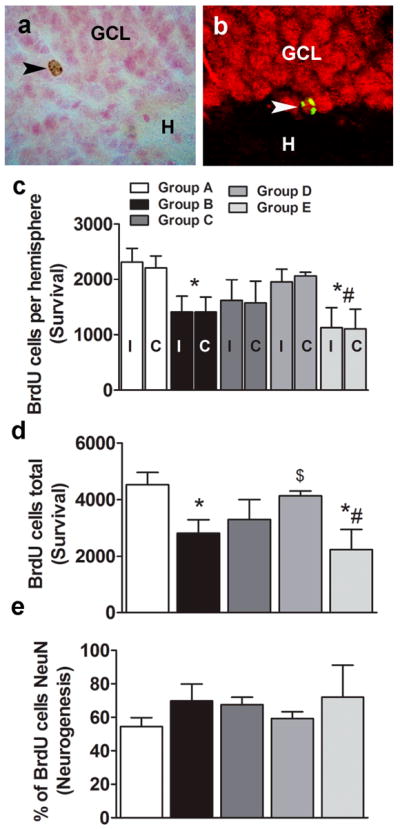
Effects of cocaine self-administration, extinction learning, and LFS during extinction learning on dentate gyrus neurogenesis. Photomicrograph of DAB-labeled (a) or fluorescent labeled, single z-stack image (BrdU, CY2, green; NeuN, CY3, red) (b) mature BrdU cell in the granule cell layer of the hippocampal dentate gyrus labeled with a BrdU antibody. Images presented are at 400x magnification. BrdU-labeled 20- to 24-day-old surviving cells are visible as oval-to-round-shaped cells. BrdU-labeled cells colocalized with NeuN cells, suggesting maturation and differentiation into granule cell neurons. GCL, granule cell layer; H, hilus. Images are 400× magnification. (c-e) Quantitative analysis of BrdU cell counts from cocaine-naive and cocaine rats from serial coronal sections. Total number of BrdU cells per hemisphere ipsilateral (I) or contralateral (C) to the LFS site (c) or combined (d). Ratio of BrdU-labeled cells colabeled with NeuN analyzed by confocal analyses (e). n = 4–6 each group. Data are expressed as mean ± SEM. *p < 0.05, compared with Group A (control); #p < 0.05, compared with Group D (non-LFS group); $p < 0.05, compared with Group B (continued cocaine group).