
Figure 4.
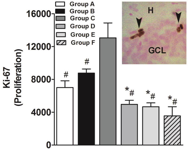
Effects of cocaine self-administration, extinction, and LFS during extinction on cell proliferation. (Main panel) Quantitative analysis of the number of DAB-labeled Ki-67-immunoreactive cells in the subgranular zone of the dentate gyrus of the hippocampus. (Inset; image presented is at 400x magnification) Photomicrograph of DAB-labeled Ki-67-immunoreactive cells. GCL, granule cell layer; H, hilus. Image is 400× magnification. n = 4–6 each group. Data are expressed as mean ± SEM. *p < 0.05, compared with Group B; #p < 0.05, compared with Group C.