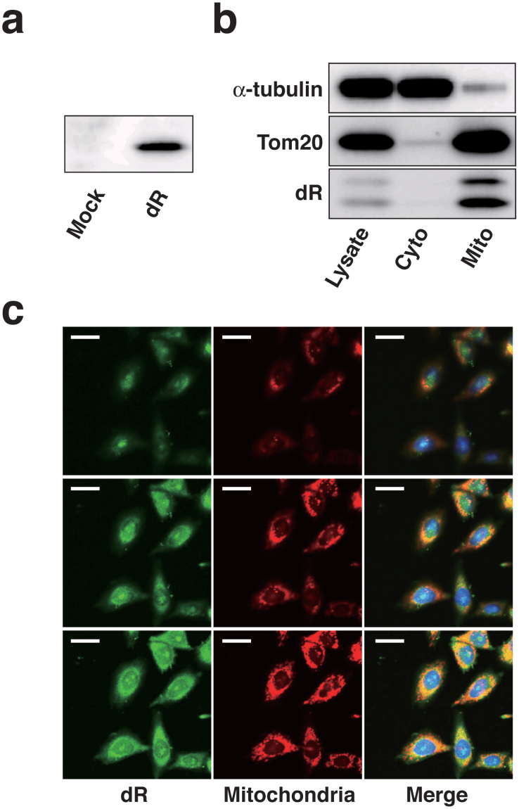
Figure 1. Mitochondrial-specific expression of dR in CHO-K1 cells.
(a) The expression of dR was confirmed using Western blotting. The 26-kDa band was observed in dR transfectants. (b) Western blot analysis after subcellular fractionations of dR expressed CHO-K1 cells. The dR protein was fractionated to mitochondria fraction, same as outer mitochondrial membrane protein, Tom20. (c) Delta-rhodopsin-pCMV/myc/mito vector was transfected into CHO-K1 cells. Z-stack confocal analysis of these cells for MitoTracker Red CMXRos and myc tag immunofluorescence were performed and merged images were indicated. Z-stacks were taken in steps of micrometer. The top to middle sections of the Z-stacks are shown. Scale bar, 20 μm. Exogenous delta-rhodopsin was co-stained with MitoTracker Red CMXRos in CHO-K1 cells.

