



Abstract
Stem cells embody the tremendous potential of the human body to develop, grow, and repair throughout life. Understanding the biologic mechanisms that underlie stem cell-mediated tissue regeneration is key to harnessing this potential. Recent advances in molecular biology, genetic engineering, and material science have broadened our understanding of stem cells and helped bring them closer to widespread clinical application. Specifically, innovative approaches to optimize how stem cells are identified, isolated, grown, and utilized will help translate these advances into effective clinical therapies. Although there is growing interest in stem cells worldwide, this enthusiasm must be tempered by the fact that these treatments remain for the most part clinically unproven. Future challenges include refining the therapeutic manipulation of stem cells, validating these technologies in randomized clinical trials, and regulating the global expansion of regenerative stem cell therapies.
Keywords: stem cell, biotechnology, regenerative medicine, tissue engineering
1. Introduction
Stem cells, which possess the ability to self-renew and differentiate into tissue-specific cells with specialized functions, have the potential to revolutionize regenerative medicine (Moore and Lemischka, 2006). They have been identified in almost all organs of the human body and play important roles in development, homeostasis, and disease (Glotzbach et al. , 2011b, Hipp and Atala, 2008). Several major classes of stem cells have been described, including embryonic stem cells (derived from pre-implantation embryos), tissue-resident stem cells (obtained from adult tissues), and induced pluripotent stem cells (iPSCs) (generated via genetic reprogramming). Given the role of stem cells in development, they have been touted as a “fountain of youth,” representing the immense potential of regenerative medicine to eradicate human suffering (te Riele, 2009).
Stem cell therapies are actively being investigated for diseases as disparate as diabetes, cancer, HIV, and wound healing (Razvi, 2012). Two clinical problems that have benefitted from stem cell therapy include hematologic cancers and severe burn injury. Transplantation of hematopoietic stem cells from the bone marrow has been extensively used to restore ablated blood cell populations after chemotherapy and may have a role in treating certain autoimmune diseases (Li and Sykes, 2012). Additionally, epithelial stem cells harvested from skin biopsies can be commercially expanded to create autologous skin equivalents used for coverage of extensive burn wounds (Atiyeh and Costagliola, 2007). These therapies highlight the potential of stem cell-based approaches to combat human suffering. Broadly speaking, regenerative medicine aims to exploit this intrinsic potential of stem cells to restore or establish normal biologic function in complex multicellular systems (Mason and Dunnill, 2008).
2. Translational challenges
Preclinical studies demonstrating the therapeutic potential of stem cells and anecdotal reports touting impressive “cures” are common in the popular media. As a result, unproven procedures using autologous stem cells are increasingly being performed worldwide, even in countries with high medical standards such as the United States (Cyranoski, 2012). Despite widespread optimism, researchers still do not fully understand the fundamental mechanisms underlying stem cell biology or how they might impact human disease (Cyranoski, 2010). Stem cells are often defined by their function in vitro, results which may not always be relevant in vivo (Prockop, 2007). Additionally, although animal studies employing stem cells have demonstrated numerous successes, these models often oversimplify human disease and may be of limited clinical applicability (Pound et al. , 2004). The ongoing challenge for clinicians and scientists is to translate findings from the laboratory into safe and effective treatments in the real world.
One potential reason why stem cell therapies have failed to impact human disease is that our concept of stem cells is loosely defined and highly imprecise (Tajbakhsh, 2009). There is little to no consensus on how different specific stem cell populations should be harvested, identified, and utilized (Tajbakhsh, 2009). Further, stem cells have been obtained from a multitude of tissues and animal species potentially accounting for the wide diversity in therapeutic outcomes. For example, researchers examining adipose-derived stem cells from mice, dogs, and humans reported dramatic differences in proliferation and osteogenic capacity (Levi et al. , 2011a). Important functional differences may even exist within stem cell populations from the same patient and tissue type (Maddox et al. , 2012, Vaculik et al. , 2012), underscoring the need to better understand what stem cell populations are actually being used.
Traditional means of defining and isolating stem cells are often based on profiles of cell surface markers that are determined empirically and/or rudimentary cell culture techniques that have changed little in decades. For example, bone marrow-derived mesenchymal stem cells are often isolated based on their adherence to culture dish plastic (Pittenger et al. , 1999). Despite the marked heterogeneity of cell populations isolated using this technique (Phinney et al. , 1999), it remains the most widely utilized method to select mesenchymal stem cells for translational studies. Given all that remains unclear about stem cell biology, it should come as no surprise that the vast majority of stem cell therapies have failed to deliver clinically consistent or robust results (Baker, 2011).
Another reason for the failures of early generation stem cell-based treatments may be related to the concept of a stem cell niche (Becerra et al. , 2011). The niche is defined as the dynamic microenvironment of biochemical and physical cues that regulate stem cell function (Li and Xie, 2005, Moore and Lemischka, 2006). In vivo, stem cells exist in tightly controlled niches that are critical in maintaining stem cell survival, differentiation, and activity. Disruption of this niche through injury, disease, or experimental manipulation can profoundly impact the ability of tissues and their constituent stem cells to repair and regenerate (Voog and Jones, 2010). However, it is important to realize that many of the diseases for which stem cells have been proposed as potential “cures” are not inherently diseases of either stem cells or the stem cell niche. In this concise review, we highlight several novel approaches our laboratory and others are pursuing to optimize stem cells for regenerative medicine.
3. Optimizing stem cell harvest and isolation
A full review of stem cell harvesting protocols and systems is beyond the scope of this review, but several novel strategies have been described. The United States Food and Drug Administration has recently approved of the use of a CXCR4 antagonist to improve hematopoietic stem cell mobilization and collection (Mahaseth and Kaufman, 2012), highlighting the potential of chemokine modulation strategies to control stem cell trafficking. Anti-adhesion molecules and anti-sense oligonucleotides have also been utilized to alter circulating stem cell and endothelial layer interactions, thus preventing stem cell egress from the circulation and improving harvest yields via the bloodstream (Kronenwett et al. , 2000).
Another approach exploits the use of computer-based harvesting. Traditional methods of stem cell isolation often entail tedious steps including manual picking of colonies and multiple subcultures. Advances in cell culture systems have enabled the development of more efficient means to harvest and maintain stem cell populations. For over a decade, peripheral blood stem cells have been effectively harvested using automated cell separator systems, with applications ranging from therapeutic plasma exchange to cell therapy for hematologic malignancies (Heuft et al. , 2000). More recently, an automated harvesting system has been shown to identify and isolate stem cell colonies efficiently (Haupt et al. , 2012). Despite these improved means to collect cells, a significant challenge remains in determining which cells are the therapeutically important cells.
Stem cells can be identified in part by their cell surface markers, a useful property for prospective isolation using flow cytometry. These surface antigen profiles are not entirely specific for specialized cell types and are based on the assumption that proteins expressed on the outside of stem cells accurately reflect their functional capacity (regulated by genes on the inside). Moreover, recent empiric studies suggest that stem cell populations are highly heterogeneous, a finding that has important implications for their differentiation potential and translational utility (Graf and Stadtfeld, 2008, Muller-Sieburg et al. , 2012, Phinney, 2012, Raaijmakers and Scadden, 2008). To address this challenge, our laboratory has developed a strategy combining microfluidic-based single cell analysis with information theory in the hopes of defining and isolating novel stem cell subgroups with different therapeutic potentials (Glotzbach et al. , 2011a).
This approach allows simultaneous gene expression analysis of up to 96 genes across hundreds of individual cells with the help of a microfluidic chip-based system (Kalisky et al. , 2011, Kalisky and Quake, 2011, Sanchez-Freire et al. , 2012). Microfluidic chips, designed to overcome the practical limitations of performing a high number of PCR reactions simultaneously, are capable of performing approximately 10,000 individual reactions on a single chip run. This high throughput technology allows for a comprehensive characterization of single cell transcriptomes, particularly useful in selecting novel stem cell populations (Figure 1). In order to utilize this information to prospectively identify and isolate highly enriched stem cell subsets within homogeneous cell populations, we developed an algorithm based on fuzzy set theory to cluster cell populations into subsets based on common gene expression signatures (Glotzbach, Januszyk, 2011a).
Figure 1. Schematic of microfluidic chip-based single cell transcriptional analysis.
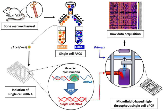
Single cells are sorted by fluorescence-activated cell sorting into individual wells of a 96-well plate that has been pre-loaded with reverse transcriptase polymerase chain reaction reagents. cDNA is created for each gene target within each individual cell followed by an initial pre-amplification step. Single cell cDNA and primer-probe sets are then loaded onto the microfluidic chip. Transcriptional analysis is performed for each cell across up to 96 genes in parallel, producing approximately 10,000 unique data points for each chip run. Figure reproduced with permission from (Glotzbach, Januszyk, 2011a).
We have utilized this approach to study a well-defined and putatively homogeneous stem cell population, murine long-term hematopoietic stem cells. We were able to demonstrate functionally unique subpopulations based on the expression of the surface marker CD34 (Glotzbach et al, 2011a) (Figure 2). In another study, we employed this technology to identify a subset of human adipose-derived stem cells with a transcriptional signature for osteogenic differentiation. Prospective isolation using negative selection for the cell surface receptor endoglin (CD105) yielded cells with dramatically increased potential for bone regeneration (Levi et al. , 2011b). Other groups have utilized similar approaches to identify transcriptional differences between iPSCs and embryonic stem cells using a selected panel of pluripotency related genes. Interestingly, iPSCs displayed higher cell-to-cell variability in the expression of pluripotency factors such as Oct4, Sox2 and Nanog, a finding that would be missed using cell population-based microarray analyses but might be of substantial therapeutic importance (Narsinh et al. , 2011).
Figure 2. Identification of distinct subgroups within a putatively homogeneous stem cell population.

(A) Hierarchical clustering of 43 genes among 300 individual cells selected for low expression of CD34, a marker of hematopoietic stem cells. Gene expression is represented as fold change from the median on a color scale from yellow (high expression) to blue (low expression). Rows = gene targets, columns = individual cells. (B) Nine genes were identified that exhibit significantly different transcriptional patterns on single cell expression between populations of high expressing and low expressing CD34 hematopoietic stem cells. (C) Comparison of these subpopulations based on hierarchical clustering of the nine genes. Note the differential expression patterns of the two stem cell subpopulations based on CD34 expression. Figure reproduced with permission from (Glotzbach, Januszyk, 2011a).
4. Improving stem cell “stemness”
Techniques have also been described to improve the stemness of cells. Ever since iPSCs were first derived via the introduction of four genes (Yamanaka factors) into somatic cells, scientists have understood that all cells contain the genetic material necessary for the development of the entire organism (Takahashi and Yamanaka, 2006). Thus, experimental manipulation of “master” transcription factors has enabled researchers to control the regenerative potential of harvested cells. For example, it has recently been shown that resident cardiac fibroblasts can be transdifferentiated into specialized cardiomyocyte-like cells in situ (Qian et al. , 2012), potentially obviating the need for cell culture-based derivation of iPSCs. Direct retroviral injection of the transcription factors Gata4, Mef2c and Tbx5 into murine hearts reprogrammed cardiac fibroblasts into cardiomyocytes and improved long-term cardiac function in a myocardial infarct model (Qian, Huang, 2012).
Additional strategies to enhance stem cell function involve exogenous manipulation via transgene technologies. Mesenchymal stem cells have been engineered to overexpress the anti-apoptosis gene Bcl-2, resulting in improved infarct recovery following intracardiac injection of stem cells in a rat model (Li et al. , 2007b). Similarly, stem cells have been transfected to overexpress hypoxia-regulated heme oxygenase-1, enabling improved stem cell engraftment and viability in infarcted hearts (Tang et al. , 2005). Researchers have also utilized polymeric nanoparticles to delivery vascular endothelial growth factor to human stem cells (Yang et al. , 2010). These stem cells were injected into ischemic hindlimbs in mice and improved limb salvage and angiogenesis. Finally, overexpression of targets associated with stem cell trafficking, survival, and cytokine secretion have been successful in augmenting the regenerative effects of delivered stem cells in preclinical trials (Cheng et al. , 2008, Li et al. , 2007a, Lim et al. , 2006).
Despite the ability of transgene technologies to manipulate stem cell behavior, the use of these systems in clinical medicine is associated with significant risks. For example, retroviruses and lentiviruses used to generate iPSCs have the potential to activate intrinsic oncogenes and subsequent tumorigenesis (Herberts et al. , 2011). Small molecules and plasmids have also been utilized to generate iPSCs but the possibility of mutagenesis and other negative side effects remains (Yamanaka, 2009). Taken together, these studies highlight the need to balance potential benefits and risks of biotechnology approaches to enhance the therapeutic potency of stem cells.
5. Optimizing the stem cell niche
Stem cells removed from their native environment behave differently in artificial culture environments (Wright, 2012). This divergence potentially becomes greater as cell populations are serially passaged, possibly resulting in important functional differences that may limit their therapeutic utility (Dey et al. , 2009, Kretlow et al. , 2008). Recent advances in highly sensitive monitoring systems and nanotopography have allowed researchers to control culture conditions precisely and restore stem cell functionality (van der Sanden et al. , 2010). Moreover, high throughput screening tools may allow researchers to identify the optimal substrate biomaterials for stem cell growth, permitting further optimization of harvest yields (Yang et al. , 2009).
Cell bioreactors are complex multi-component systems that enable researchers to grow cells and tissues in controlled culture settings. These engineering devices, which recapitulate biologically active microenvironments using modifiable platforms that include stirring, perfusion, and/or microfluidic systems, have been shown to improve harvest of multiple stem cell types (Godara et al. , 2008). Neural stem cells have been successfully grown using bioreactors with perfused scaffolds and complex pH/oxygen control capabilities (King and Miller, 2007) and mouse embryonic stem cells have been efficiently differentiated into cardiomyocytes using a stirred bioreactor device (Schroeder et al. , 2005). Bioreactors will likely play an important role in the development of stem cell therapies by integrating traditional biochemical/physical engineering processes with novel cell biology technologies to scale up stem cell culture systems for clinical use (Rodrigues et al. , 2011, Sharma et al. , 2011).
Three-dimensional culture systems have enabled higher cell yields compared to conventional two-dimensional methods (Justice et al. , 2009). Thus, traditional brightfield microscopic analysis of biologic parameters including confluency, morphology, and proliferation may not be applicable for bioreactor-based cultures. For example, fluorescent cell markers have been used to assess neuronal morphology in two versus three dimensional culture conditions, demonstrating important quantitative differences in differentiation (Arien-Zakay et al. , 2009). Novel techniques to monitor cell growth and quality have also been developed using multidimensional image analysis (Baradez and Marshall, 2011). As stem cell research moves from the laboratory into the clinics, more efficient and precise systems will be needed for large-scale manufacturing of high-grade therapeutic stem cells.
Another major challenge for stem cell therapy is how to maintain the regenerative potential of stem cells during and after delivery. We have previously proposed the concept of “seed vs. soil” to illustrate the importance of the microenvironment in regenerative processes (Gurtner et al. , 2008). Stem cells are highly regulated by their niche (the local biochemical and physical cues from surrounding matrix and cells that drive stemness), which is disrupted immediately following cell harvest (Moore and Lemischka, 2006, Voog and Jones, 2010). Further, dynamic interactions between stem cells, differentiating progeny, and local differentiated cells may maintain niche organization, suggesting that delivery of homogeneous stem cell populations alone may be inadequate for regenerative purposes (Scadden, 2006). Although stem cells maintain the ability to differentiate and self-renew under highly controlled culture conditions, it is unclear how much of this intrinsic regenerative potential is lost ex vivo. This problem may be further exacerbated by cell delivery systems that fail to account for the complex cellular, structural, and soluble networks that comprise the stem cell niche.
Stem cells can be therapeutically delivered via various approaches, including topical, injection, and systemic delivery (Figure 3). Topical delivery of stem cells is not possible for deeper tissues and organs but may have a role for cutaneous wounds or during surgical reconstruction where tissue defects can be directly accessed. Injection methods involve the mass delivery of cells into tight spaces under high pressures, conditions which are likely unfavorable for cell engraftment and growth. Systemic delivery relies on stem cells being able to effectively traffic and egress to the injury site. For each of these approaches, stem cells must re-establish their niche environment in order to both self-renew and differentiate during tissue regeneration.
Figure 3. Stem cell delivery methods.

Commonly used methods to delivery stem cells include topical, injection, systemic, or scaffold-mediated approaches. Topical strategies are limited to sites that can be easily accessed, including cutaneous wounds or during surgical procedures. Injection therapies can access most anatomic sites, but stem cells are often delivered under hostile conditions (high pressures and cell densities). Systemic strategies require the ability of stem cells to properly home to damaged tissues and to effectively egress the circulation and enter the interstitial space. Scaffold-based techniques appear to be highly promising, offering the potential of a protective, three-dimensional vehicle with controlled spatial cues and precisely patterned matrix and cytokine gradients.
Most preclinical and clinical studies have failed to demonstrate prolonged survival of stem cells following delivery despite observed therapeutic benefits (Prockop, 2007). Many groups have attributed positive outcomes to stem cell secretion of paracrine signals as opposed to long-term engraftment and differentiation (Rustad et al. , 2012, Wu et al. , 2007), leading one to question whether stem cell-mediated regeneration is actually occurring. Clinical experience using stem cells has likewise been promising but limited to small case reports and non-controlled studies that have not evaluated long-term engraftment (Dash et al. , 2009, Macchiarini et al. , 2008, Renner et al. , 2009, Rigotti et al. , 2007). Thus, whether therapeutic stem cells contribute to tissue regeneration directly via tissue-specific differentiation or whether they act as temporary cytokine vehicles remains unclear.
One strategy to improve the clinical utility of stem cells is the use of scaffolds. Both naturally derived and synthetic scaffolds have been used successfully in conjunction with stem cells for tissue engineering (Chaikof et al. , 2002, Chan and Leong, 2008, Dawson et al. , 2008, Tateishi et al. , 2002, Williams, 2009). This approach enables the delivery of cells in a structured matrix environment that provides protection and controlled spatial cues in a non-native setting (Figure 3). The matrix components themselves can be modified to regulate stem cell function on increasingly precise scales. Additionally, numerous studies have shown that control release systems can be incorporated into scaffolds to modulate stem cell activity further via growth factors, small molecules, plasmids, or other vectors (Ioannidou, 2006). A full review of scaffolds and cytokines for stem cell therapy is beyond the scope of this review, but we refer the reader to several excellent papers on this topic (Chan and Leong, 2008, Dawson, Mapili, 2008, Lee et al. , 2011, Ma, 2008, Place et al. , 2009, Salgado et al. , 2010).
Another approach utilizes bioengineered scaffolds that actively modify their surroundings to enhance stem cell incorporation. Our laboratory has focused on developing stem cell therapies for acute wounds that often produce high levels of damaging free radicals. We have previously developed a carbohydrate-based hydrogel scaffold that promotes neovascularization in cutaneous wounds (Wong et al. , 2011a) and enhances the stemness of mesenchymal stem cells (Rustad, Wong, 2012) (Figures 4 and 5). This engineered hydrogel also exhibits potent anti-oxidant properties that were shown to improve stem cell engraftment in ischemic wounds compared to injection delivery, results attributed to quenching of free radicals by the biomaterial itself (Wong et al. , 2011b).
Figure 4. Biomaterial-enhanced expression of stemness genes in mesenchymal stem cells.

(A) Reverse transcriptase polymerase chain reaction analysis of Oct4, Sox2, and Klf4 expression in mouse mesenchymal stem cells seeded into a carbohydrate-based dermal hydrogel compared to standard tissue culture plate. (B) Immunoblot protein validation and (C) quantification. (D) Immunofluorescence imaging demonstrates increased expression of stemness markers with seeding into a dermal hydrogel, suggesting enhancement of cell stemness via a biomaterial niche-driven approach. *p<0.05. Figure reproduced with permission from (Rustad, Wong, 2012).
Figure 5. Commercial development of a dermal hydrogel for stem cell-mediated wound healing.

(A) Based on promising preclinical results, our laboratory is developing a dry hydrogel dressing that can be readily seeded with stem cells in solution. (B) Stem cells engrafted throughout the dermal-like matrix can be delivered directly into cutaneous wounds in a niche-like environment that has been demonstrated to promote wound vascularization and stem cell potency. (C) A topical adhesive covering is being developed to protect and secure the dermal hydrogel.
Collectively, these strategies demonstrate the tremendous promise of biotechnology to advance stem cell therapies for human disease. By building on the fundamental units of tissue engineering (cells, matrices, and growth factors) (Langer and Vacanti, 1993), researchers will continue to develop multidimensional systems capable of exploiting the ability of progenitor cells to promote tissue repair. The concept of creating or recapitulating a “therapeutic niche” will play an increasingly important role in understanding how to harness the regenerative potential of stem cells.
6. Future directions
Amphibians and simpler organisms demonstrate the ability to restore complex tissues/organs following injury and have served as useful models to study regenerative processes. Mammalian regenerative capabilities may not be as impressive, but organs such as the human liver can re-grow following significant injury. Moreover, human fetal wounds have been shown to heal without scar and reports of digit regeneration following amputation indicate that mammals have maintained some degree of tissue regeneration (Gurtner, Werner, 2008). Recent studies using a digit amputation model in transgenic mice have shown that lineage-specific, germ layer-restricted stem cells present at the injury site are sufficient to regenerate highly complex tissues (Rinkevich et al. , 2011). Future studies will be needed to understand further the importance of molecular and stem cell-associated pathways in adult regeneration.
Another major challenge is how to validate regenerative stem cell therapies. Rodent models serve as a practical tool to explore investigational therapies and molecular mechanisms but their value in advancing clinical treatments remains controversial (Ferdowsian and Beck, 2011, Pound, Ebrahim, 2004). Animal models in dogs, sheep, pigs, and non-human primates may provide greater insight into the applicability of experimental stem cell therapies. Recent reviews that detail the use of cardiac, hematopoietic, and neurologic stem cells in large animal models highlight their growing relevance in translating pre-clinical stem cell technologies (Gandolfi et al. , 2011, Klassen et al. , 2012, Trobridge and Kiem, 2010).
Successes in the research laboratory are often promoted as miracle cures by the press, driving public interest in stem cell treatments and creating a growing global industry referred to as “stem cell tourism” (Einsiedel and Adamson, 2012). As yet, this explosion of stem cell therapies worldwide has not been followed by clinical trials demonstrating their safety or efficacy. International guidelines such as those published by the International Society for Stem Cell Research are an important step in ensuring that the translational use of stem cells meets rigorous “scientific, clinical, regulatory, ethical, and societal” standards (2008). The regulation of this emerging technology needs to be balanced with continued support of innovation to ultimately impact global health.
The stem cell therapy industry is maturing into a viable environment for investment with annual revenues estimated at over $1 billion US (Mason et al. , 2011). Although initial funding for stem cell companies has typically been provided by angel and venture capital investment, clinical translation often requires additional funding that is difficult to obtain. The high volatility of this sector has further limited the availability of sustainable sources of investment (Brindley et al. , 2011). However recently, publically-funded centers associated with academic institutions have emerged to help bridge translational cell therapies in a responsible manner (Mason, Brindley, 2011). The future stability and growth of this industry will depend ultimately on scalable and cost-efficient strategies that lead to clinical therapies. Despite these and other ongoing challenges, regenerative stem cell treatments based on biotechnological innovation have the potential to revolutionize modern medicine.
Acknowledgments
Funding for stem cell research conducted in our laboratory has been provided by the Hagey Family Endowed Fund in Stem Cell Research and Regenerative Medicine, the Armed Forces Institute of Regenerative Medicine (United States Department of Defense), the National Institutes of Health (R01-DK074095 and R01-AG025016), and the Oak Foundation. The authors would also like to thank the following researchers for their contributions to the data presented in this paper: Jason P. Glotzbach, Michael Januszyk, Ivan N. Vial, Kristine C. Rustad, and Jayakumar Rajadas.
Glossary
- n/a
Non-standard abbreviations
Footnotes
Publisher's Disclaimer: This is a PDF file of an unedited manuscript that has been accepted for publication. As a service to our customers we are providing this early version of the manuscript. The manuscript will undergo copyediting, typesetting, and review of the resulting proof before it is published in its final citable form. Please note that during the production process errors may be discovered which could affect the content, and all legal disclaimers that apply to the journal pertain.
References
- Guidelines for the Clinical Treatment of Stem Cells. International Society for Stem Cell Research Task Force. 2008 [Google Scholar]
- Arien-Zakay H, Lecht S, Perets A, Roszell B, Lelkes PI, Lazarovici P. Quantitative assessment of neuronal differentiation in three-dimensional collagen gels using enhanced green fluorescence protein expressing PC12 pheochromocytoma cells. J Mol Neurosci. 2009;37:225–37. doi: 10.1007/s12031-008-9123-1. [DOI] [PubMed] [Google Scholar]
- Atiyeh BS, Costagliola M. Cultured epithelial autograft (CEA) in burn treatment: three decades later. Burns. 2007;33:405–13. doi: 10.1016/j.burns.2006.11.002. [DOI] [PubMed] [Google Scholar]
- Baker M. Stem-cell pioneer bows out. Nature. 2011;479:459. doi: 10.1038/479459a. [DOI] [PubMed] [Google Scholar]
- Baradez MO, Marshall D. The use of multidimensional image-based analysis to accurately monitor cell growth in 3D bioreactor culture. PLoS One. 2011;6:e26104. doi: 10.1371/journal.pone.0026104. [DOI] [PMC free article] [PubMed] [Google Scholar]
- Becerra J, Santos-Ruiz L, Andrades JA, Mari-Beffa M. The stem cell niche should be a key issue for cell therapy in regenerative medicine. Stem Cell Rev. 2011;7:248–55. doi: 10.1007/s12015-010-9195-5. [DOI] [PubMed] [Google Scholar]
- Brindley DA, Reeve BC, Sahlman WA, Bonfiglio GA, Davie NL, Culme-Seymour EJ, et al. The impact of market volatility on the cell therapy industry. Cell Stem Cell. 2011;9:397–401. doi: 10.1016/j.stem.2011.10.010. [DOI] [PubMed] [Google Scholar]
- Chaikof E, Matthew H, Kohn J, Mikos A, Prestwich G, Yip C. Biomaterials and scaffolds in reparative medicine. Annals of the New York Academy of Sciences. 2002;961:96–105. doi: 10.1111/j.1749-6632.2002.tb03057.x. [DOI] [PubMed] [Google Scholar]
- Chan B, Leong K. Scaffolding in tissue engineering: general approaches and tissue-specific considerations. European Spine Journal. 2008;17:467–79. doi: 10.1007/s00586-008-0745-3. [DOI] [PMC free article] [PubMed] [Google Scholar]
- Cheng Z, Ou L, Zhou X, Li F, Jia X, Zhang Y, et al. Targeted migration of mesenchymal stem cells modified with CXCR4 gene to infarcted myocardium improves cardiac performance. Mol Ther. 2008;16:571–9. doi: 10.1038/sj.mt.6300374. [DOI] [PubMed] [Google Scholar]
- Cyranoski D. Strange lesions after stem-cell therapy. Nature. 2010;465:997. doi: 10.1038/465997a. [DOI] [PubMed] [Google Scholar]
- Cyranoski D. Stem-cell therapy takes off in Texas. Nature. 2012;483:13–4. doi: 10.1038/483013a. [DOI] [PubMed] [Google Scholar]
- Dash N, Dash S, Routray P, Mohapatra S, Mohapatra P. Targeting nonhealing ulcers of lower extremity in human through autologous bone marrow-derived mesenchymal stem cells. Rejuvenation Research. 2009;12:359–66. doi: 10.1089/rej.2009.0872. [DOI] [PubMed] [Google Scholar]
- Dawson E, Mapili G, Erickson K, Taqvi S, Roy K. Biomaterials for stem cell differentiation. Adv Drug Deliv Rev. 2008;60:215–28. doi: 10.1016/j.addr.2007.08.037. [DOI] [PubMed] [Google Scholar]
- Dey D, Saxena M, Paranjape AN, Krishnan V, Giraddi R, Kumar MV, et al. Phenotypic and functional characterization of human mammary stem/progenitor cells in long term culture. PLoS One. 2009;4:e5329. doi: 10.1371/journal.pone.0005329. [DOI] [PMC free article] [PubMed] [Google Scholar]
- Einsiedel EF, Adamson H. Stem cell tourism and future stem cell tourists: policy and ethical implications. Dev World Bioeth. 2012;12:35–44. doi: 10.1111/j.1471-8847.2012.00319.x. [DOI] [PubMed] [Google Scholar]
- Ferdowsian HR, Beck N. Ethical and scientific considerations regarding animal testing and research. PLoS One. 2011;6:e24059. doi: 10.1371/journal.pone.0024059. [DOI] [PMC free article] [PubMed] [Google Scholar]
- Gandolfi F, Vanelli A, Pennarossa G, Rahaman M, Acocella F, Brevini TA. Large animal models for cardiac stem cell therapies. Theriogenology. 2011;75:1416–25. doi: 10.1016/j.theriogenology.2011.01.026. [DOI] [PubMed] [Google Scholar]
- Glotzbach JP, Januszyk M, Vial IN, Wong VW, Gelbard A, Kalisky T, et al. An information theoretic, microfluidic-based single cell analysis permits identification of subpopulations among putatively homogeneous stem cells. PLoS One. 2011a;6:e21211. doi: 10.1371/journal.pone.0021211. [DOI] [PMC free article] [PubMed] [Google Scholar]
- Glotzbach JP, Wong VW, Gurtner GC, Longaker MT. Regenerative medicine. Curr Probl Surg. 2011b;48:148–212. doi: 10.1067/j.cpsurg.2010.11.002. [DOI] [PubMed] [Google Scholar]
- Godara P, McFarland CD, Nordon RE. Design of bioreactors for mesenchymal stem cell tissue engineering. J Chem Technol Biotechnol. 2008;83:408–20. [Google Scholar]
- Graf T, Stadtfeld M. Heterogeneity of embryonic and adult stem cells. Cell Stem Cell. 2008;3:480–3. doi: 10.1016/j.stem.2008.10.007. [DOI] [PubMed] [Google Scholar]
- Gurtner GC, Werner S, Barrandon Y, Longaker MT. Wound repair and regeneration. Nature. 2008;453:314–21. doi: 10.1038/nature07039. [DOI] [PubMed] [Google Scholar]
- Haupt S, Grützner J, Thier MC, Kallweit T, Rath BH, Laufenberg I, et al. Automated selection and harvesting of pluripotent stem cell colonies. Biotechnol Appl Biochem. 2012;59:77–87. doi: 10.1002/bab.1014. [DOI] [PubMed] [Google Scholar]
- Herberts CA, Kwa MS, Hermsen HP. Risk factors in the development of stem cell therapy. J Transl Med. 2011;9:29. doi: 10.1186/1479-5876-9-29. [DOI] [PMC free article] [PubMed] [Google Scholar]
- Heuft HG, Dubiel M, Kingreen D, Oertel J, De Reys S, Rick O, et al. Automated collection of peripheral blood stem cells with the COBE spectra for autotransplantation. Vox Sang. 2000;79:94–9. doi: 10.1159/000031219. [DOI] [PubMed] [Google Scholar]
- Hipp J, Atala A. Sources of stem cells for regenerative medicine. Stem Cell Rev. 2008;4:3–11. doi: 10.1007/s12015-008-9010-8. [DOI] [PubMed] [Google Scholar]
- Ioannidou E. Therapeutic modulation of growth factors and cytokines in regenerative medicine. Curr Pharm Des. 2006;12:2397–408. doi: 10.2174/138161206777699007. [DOI] [PubMed] [Google Scholar]
- Justice BA, Badr NA, Felder RA. 3D cell culture opens new dimensions in cell-based assays. Drug Discov Today. 2009;14:102–7. doi: 10.1016/j.drudis.2008.11.006. [DOI] [PubMed] [Google Scholar]
- Kalisky T, Blainey P, Quake SR. Genomic analysis at the single-cell level. Annu Rev Genet. 2011;45:431–45. doi: 10.1146/annurev-genet-102209-163607. [DOI] [PMC free article] [PubMed] [Google Scholar]
- Kalisky T, Quake SR. Single-cell genomics. Nat Methods. 2011;8:311–4. doi: 10.1038/nmeth0411-311. [DOI] [PubMed] [Google Scholar]
- King JA, Miller WM. Bioreactor development for stem cell expansion and controlled differentiation. Curr Opin Chem Biol. 2007;11:394–8. doi: 10.1016/j.cbpa.2007.05.034. [DOI] [PMC free article] [PubMed] [Google Scholar]
- Klassen H, Tucker BA, Liew CG, La Cour M, Wang HC. Stem cells in large animal models of retinal and neurological disease. Stem Cells Int. 2012;2012:460504. doi: 10.1155/2012/460504. [DOI] [PMC free article] [PubMed] [Google Scholar]
- Kretlow JD, Jin YQ, Liu W, Zhang WJ, Hong TH, Zhou G, et al. Donor age and cell passage affects differentiation potential of murine bone marrow-derived stem cells. BMC Cell Biol. 2008;9:60. doi: 10.1186/1471-2121-9-60. [DOI] [PMC free article] [PubMed] [Google Scholar]
- Kronenwett R, Martin S, Haas R. The role of cytokines and adhesion molecules for mobilization of peripheral blood stem cells. Stem Cells. 2000;18:320–30. doi: 10.1634/stemcells.18-5-320. [DOI] [PubMed] [Google Scholar]
- Langer R, Vacanti JP. Tissue engineering. Science. 1993;260:920–6. doi: 10.1126/science.8493529. [DOI] [PubMed] [Google Scholar]
- Lee K, Silva EA, Mooney DJ. Growth factor delivery-based tissue engineering: general approaches and a review of recent developments. J R Soc Interface. 2011;8:153–70. doi: 10.1098/rsif.2010.0223. [DOI] [PMC free article] [PubMed] [Google Scholar]
- Levi B, Nelson ER, Brown K, James AW, Xu D, Dunlevie R, et al. Differences in osteogenic differentiation of adipose-derived stromal cells from murine, canine, and human sources in vitro and in vivo. Plast Reconstr Surg. 2011a;128:373–86. doi: 10.1097/PRS.0b013e31821e6e49. [DOI] [PMC free article] [PubMed] [Google Scholar]
- Levi B, Wan DC, Glotzbach JP, Hyun J, Januszyk M, Montoro D, et al. CD105 protein depletion enhances human adipose-derived stromal cell osteogenesis through reduction of transforming growth factor beta1 (TGF-beta1) signaling. J Biol Chem. 2011b;286:39497–509. doi: 10.1074/jbc.M111.256529. [DOI] [PMC free article] [PubMed] [Google Scholar]
- Li H, Dai K, Tang T, Zhang X, Yan M, Lou J. Bone regeneration by implantation of adipose-derived stromal cells expressing BMP-2. Biochem Biophys Res Commun. 2007a;356:836–42. doi: 10.1016/j.bbrc.2007.02.165. [DOI] [PubMed] [Google Scholar]
- Li HW, Sykes M. Emerging concepts in haematopoietic cell transplantation. Nat Rev Immunol. 2012;12:403–16. doi: 10.1038/nri3226. [DOI] [PMC free article] [PubMed] [Google Scholar]
- Li L, Xie T. Stem cell niche: structure and function. Ann Rev Cell Dev Biol. 2005;21:605–31. doi: 10.1146/annurev.cellbio.21.012704.131525. [DOI] [PubMed] [Google Scholar]
- Li W, Ma N, Ong LL, Nesselmann C, Klopsch C, Ladilov Y, et al. Bcl-2 engineered MSCs inhibited apoptosis and improved heart function. Stem Cells. 2007b;25:2118–27. doi: 10.1634/stemcells.2006-0771. [DOI] [PubMed] [Google Scholar]
- Lim SY, Kim YS, Ahn Y, Jeong MH, Hong MH, Joo SY, et al. The effects of mesenchymal stem cells transduced with Akt in a porcine myocardial infarction model. Cardiovasc Res. 2006;70:530–42. doi: 10.1016/j.cardiores.2006.02.016. [DOI] [PubMed] [Google Scholar]
- Ma PX. Biomimetic materials for tissue engineering. Adv Drug Deliv Rev. 2008;60:184–98. doi: 10.1016/j.addr.2007.08.041. [DOI] [PMC free article] [PubMed] [Google Scholar]
- Macchiarini P, Jungebluth P, Go T, Asnaghi MA, Rees LE, Cogan TA, et al. Clinical transplantation of a tissue-engineered airway. The Lancet. 2008;372:2023–30. doi: 10.1016/S0140-6736(08)61598-6. [DOI] [PubMed] [Google Scholar]
- Maddox JR, Ludlow KD, Li F, Niyibizi C. Breast and abdominal adipose multipotent mesenchymal stromal cells and stage-specific embryonic antigen 4 expression. Cells Tissues Organs. 2012;196:107–16. doi: 10.1159/000331332. [DOI] [PMC free article] [PubMed] [Google Scholar]
- Mahaseth H, Kaufman J. Optimizing stem cell collection through CXCR4 antagonists. Front Biosci (Schol Ed) 2012;4:611–9. doi: 10.2741/s288. [DOI] [PubMed] [Google Scholar]
- Mason C, Brindley DA, Culme-Seymour EJ, Davie NL. Cell therapy industry: billion dollar global business with unlimited potential. Regen Med. 2011;6:265–72. doi: 10.2217/rme.11.28. [DOI] [PubMed] [Google Scholar]
- Mason C, Dunnill P. A brief definition of regenerative medicine. Regen Med. 2008;3:1–5. doi: 10.2217/17460751.3.1.1. [DOI] [PubMed] [Google Scholar]
- Moore KA, Lemischka IR. Stem cells and their niches. Science. 2006;311:1880–5. doi: 10.1126/science.1110542. [DOI] [PubMed] [Google Scholar]
- Muller-Sieburg CE, Sieburg HB, Bernitz JM, Cattarossi G. Stem cell heterogeneity: implications for aging and regenerative medicine. Blood. 2012;119:3900–7. doi: 10.1182/blood-2011-12-376749. [DOI] [PMC free article] [PubMed] [Google Scholar]
- Narsinh KH, Sun N, Sanchez-Freire V, Lee AS, Almeida P, Hu S, et al. Single cell transcriptional profiling reveals heterogeneity of human induced pluripotent stem cells. J Clin Invest. 2011;121:1217–21. doi: 10.1172/JCI44635. [DOI] [PMC free article] [PubMed] [Google Scholar]
- Phinney DG. Functional heterogeneity of mesenchymal stem cells: implications for cell therapy. J Cell Biochem. 2012;113:2806–12. doi: 10.1002/jcb.24166. [DOI] [PubMed] [Google Scholar]
- Phinney DG, Kopen G, Isaacson RL, Prockop DJ. Plastic adherent stromal cells from the bone marrow of commonly used strains of inbred mice: variations in yield, growth, and differentiation. J Cell Biochem. 1999;72:570–85. [PubMed] [Google Scholar]
- Pittenger MF, Mackay AM, Beck SC, Jaiswal RK, Douglas R, Mosca JD, et al. Multilineage potential of adult human mesenchymal stem cells. Science. 1999;284:143–7. doi: 10.1126/science.284.5411.143. [DOI] [PubMed] [Google Scholar]
- Place ES, George JH, Williams CK, Stevens MM. Synthetic polymer scaffolds for tissue engineering. Chem Soc Rev. 2009;38:1139–51. doi: 10.1039/b811392k. [DOI] [PubMed] [Google Scholar]
- Pound P, Ebrahim S, Sandercock P, Bracken MB, Roberts I. Where is the evidence that animal research benefits humans? BMJ. 2004;328:514–7. doi: 10.1136/bmj.328.7438.514. [DOI] [PMC free article] [PubMed] [Google Scholar]
- Prockop DJ. “Stemness” does not explain the repair of many tissues by mesenchymal stem/multipotent stromal cells (MSCs) Clin Pharmacol Ther. 2007;82:241–3. doi: 10.1038/sj.clpt.6100313. [DOI] [PubMed] [Google Scholar]
- Qian L, Huang Y, Spencer CI, Foley A, Vedantham V, Liu L, et al. In vivo reprogramming of murine cardiac fibroblasts into induced cardiomyocytes. Nature. 2012;485:593–8. doi: 10.1038/nature11044. [DOI] [PMC free article] [PubMed] [Google Scholar]
- Raaijmakers MH, Scadden DT. Divided within: heterogeneity within adult stem cell pools. Cell. 2008;135:1006–8. doi: 10.1016/j.cell.2008.11.034. [DOI] [PubMed] [Google Scholar]
- Razvi E. Conference scene: trends in the stem cells marketplace - report from Select Biosciences Stem Cells 2012 Conference. Regen Med. 2012;7:291–4. doi: 10.2217/rme.12.18. [DOI] [PubMed] [Google Scholar]
- Renner R, Harth W, Simon JC. Transplantation of chronic wounds with epidermal sheets derived from autologous hair follicles--the Leipzig experience. Int Wound J. 2009;6:226–32. doi: 10.1111/j.1742-481X.2009.00609.x. [DOI] [PMC free article] [PubMed] [Google Scholar]
- Rigotti G, Marchi A, Galie M, Baroni G, Benati D, Krampera M, et al. Clinical treatment of radiotherapy tissue damage by lipoaspirate transplant: a healing process mediated by adipose-derived adult stem cells. Plast Reconstr Surg. 2007;119:1409–22. doi: 10.1097/01.prs.0000256047.47909.71. [DOI] [PubMed] [Google Scholar]
- Rinkevich Y, Lindau P, Ueno H, Longaker MT, Weissman IL. Germ-layer and lineage-restricted stem/progenitors regenerate the mouse digit tip. Nature. 2011;476:409–13. doi: 10.1038/nature10346. [DOI] [PMC free article] [PubMed] [Google Scholar]
- Rodrigues CA, Fernandes TG, Diogo MM, da Silva CL, Cabral JM. Stem cell cultivation in bioreactors. Biotechnol Adv. 2011;29:815–29. doi: 10.1016/j.biotechadv.2011.06.009. [DOI] [PubMed] [Google Scholar]
- Rustad KC, Wong VW, Sorkin M, Glotzbach JP, Major MR, Rajadas J, et al. Enhancement of mesenchymal stem cell angiogenic capacity and stemness by a biomimetic hydrogel scaffold. Biomaterials. 2012;33:80–90. doi: 10.1016/j.biomaterials.2011.09.041. [DOI] [PMC free article] [PubMed] [Google Scholar]
- Salgado AJ, Reis RL, Sousa NJ, Gimble JM. Adipose tissue derived stem cells secretome: soluble factors and their roles in regenerative medicine. Curr Stem Cell Res Ther. 2010;5:103–10. doi: 10.2174/157488810791268564. [DOI] [PubMed] [Google Scholar]
- Sanchez-Freire V, Ebert AD, Kalisky T, Quake SR, Wu JC. Microfluidic single-cell real-time PCR for comparative analysis of gene expression patterns. Nat Protoc. 2012;7:829–38. doi: 10.1038/nprot.2012.021. [DOI] [PMC free article] [PubMed] [Google Scholar]
- Scadden DT. The stem-cell niche as an entity of action. Nature. 2006;441:1075–9. doi: 10.1038/nature04957. [DOI] [PubMed] [Google Scholar]
- Schroeder M, Niebruegge S, Werner A, Willbold E, Burg M, Ruediger M, et al. Differentiation and lineage selection of mouse embryonic stem cells in a stirred bench scale bioreactor with automated process control. Biotechnol Bioeng. 2005;92:920–33. doi: 10.1002/bit.20668. [DOI] [PubMed] [Google Scholar]
- Sharma S, Raju R, Sui S, Hu WS. Stem cell culture engineering - process scale up and beyond. Biotechnol J. 2011;6:1317–29. doi: 10.1002/biot.201000435. [DOI] [PubMed] [Google Scholar]
- Tajbakhsh S. Stem cell: what's in a name? Nat Reports Stem Cells. 2009 [Google Scholar]
- Takahashi K, Yamanaka S. Induction of pluripotent stem cells from mouse embryonic and adult fibroblast cultures by defined factors. Cell. 2006;126:663–76. doi: 10.1016/j.cell.2006.07.024. [DOI] [PubMed] [Google Scholar]
- Tang YL, Tang Y, Zhang YC, Qian K, Shen L, Phillips MI. Improved graft mesenchymal stem cell survival in ischemic heart with a hypoxia-regulated heme oxygenase-1 vector. J Am Coll Cardiol. 2005;46:1339–50. doi: 10.1016/j.jacc.2005.05.079. [DOI] [PubMed] [Google Scholar]
- Tateishi T, Chen G, Ushida T. Biodegradable porous scaffolds for tissue engineering. Journal of Artificial Organs. 2002;5:77–83. [Google Scholar]
- te Riele H. Recreating stem cells: a novel entrance to the fountain of youth. Cell Stem Cell. 2009;4:279–80. doi: 10.1016/j.stem.2009.03.011. [DOI] [PubMed] [Google Scholar]
- Trobridge GD, Kiem HP. Large animal models of hematopoietic stem cell gene therapy. Gene Ther. 2010;17:939–48. doi: 10.1038/gt.2010.47. [DOI] [PMC free article] [PubMed] [Google Scholar]
- Vaculik C, Schuster C, Bauer W, Iram N, Pfisterer K, Kramer G, et al. Human dermis harbors distinct mesenchymal stromal cell subsets. J Invest Dermatol. 2012;132:563–74. doi: 10.1038/jid.2011.355. [DOI] [PMC free article] [PubMed] [Google Scholar]
- van der Sanden B, Dhobb M, Berger F, Wion D. Optimizing stem cell culture. J Cell Biochem. 2010;111:801–7. doi: 10.1002/jcb.22847. [DOI] [PMC free article] [PubMed] [Google Scholar]
- Voog J, Jones DL. Stem cells and the niche: a dynamic duo. Cell Stem Cell. 2010;6:103–15. doi: 10.1016/j.stem.2010.01.011. [DOI] [PMC free article] [PubMed] [Google Scholar]
- Williams DF. On the nature of biomaterials. Biomaterials. 2009;30:5897–909. doi: 10.1016/j.biomaterials.2009.07.027. [DOI] [PubMed] [Google Scholar]
- Wong VW, Rustad KC, Galvez MG, Neofytou E, Glotzbach JP, Januszyk M, et al. Engineered pullulancollagen composite dermal hydrogels improve early cutaneous wound healing. Tissue Eng Part A. 2011a;17:631–44. doi: 10.1089/ten.tea.2010.0298. [DOI] [PMC free article] [PubMed] [Google Scholar]
- Wong VW, Rustad KC, Glotzbach JP, Sorkin M, Inayathullah M, Major MR, et al. Pullulan hydrogels improve mesenchymal stem cell delivery into high-oxidative-stress wounds. Macromol Biosci. 2011b;11:1458–66. doi: 10.1002/mabi.201100180. [DOI] [PMC free article] [PubMed] [Google Scholar]
- Wright NA. Stem cell identification--in vivo lineage analysis versus in vitro isolation and clonal expansion. J Pathol. 2012;227:255–66. doi: 10.1002/path.4018. [DOI] [PubMed] [Google Scholar]
- Wu Y, Chen L, Scott PG, Tredget EE. Mesenchymal stem cells enhance wound healing through differentiation and angiogenesis. Stem Cells. 2007;25:2648–59. doi: 10.1634/stemcells.2007-0226. [DOI] [PubMed] [Google Scholar]
- Yamanaka S. A fresh look at iPS cells. Cell. 2009;137:13–7. doi: 10.1016/j.cell.2009.03.034. [DOI] [PubMed] [Google Scholar]
- Yang F, Cho SW, Son SM, Bogatyrev SR, Singh D, Green JJ, et al. Genetic engineering of human stem cells for enhanced angiogenesis using biodegradable polymeric nanoparticles. Proc Natl Acad Sci U S A. 2010;107:3317–22. doi: 10.1073/pnas.0905432106. [DOI] [PMC free article] [PubMed] [Google Scholar]
- Yang F, Mei Y, Langer R, Anderson DG. High throughput optimization of stem cell microenvironments. Comb Chem High Throughput Screen. 2009;12:554–61. doi: 10.2174/138620709788681916. [DOI] [PMC free article] [PubMed] [Google Scholar]