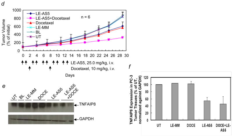
Figure 2.
Enhanced antitumor efficacy of a combination of ionizing radiation or docetaxel and systemically administered liposome-entrapped TNFAIP8 antisense oligonucleotide (LE-AS5) in PC-3 tumor xenograft model. (a) LE-AS5-mediated PC-3 tumor radiosensitization in vivo. PC-3 tumor bearing athymic mice were randomized into six groups. LE-AS5 and LE-AS5 + IR treatment groups received 25.0 mg/kg/dose (i.v., ×10) of LE-AS5 over 14 days. IR alone and LE-AS5 + IR groups received 3.8 Gy/day IR daily (day 5 through day 8). BL and LE-MM groups received blank liposomes and liposome-encapsulated mismatch oligo, respectively, at the same dosing schedule as LE-AS5. Additional control group was left untreated (UT). Tumor volumes were monitored and individual tumor volume (% initial) was calculated as the percentage of pre-treatment tumor volume (day 0, the first day of dosing; 100%). The values shown are mean tumor volume (% initial) ± S.E. (b) Inhibitory effect of LE-AS5 on TNFAIP8 expression in PC-3 tumor tissues. Two mice from each treatment group in panel (a) were sacrificed within 6–12 hr after the last dosing, and TNFAIP8 expression in tumor tissue homogenates as analyzed by Western blotting with anti-TNFAIP8 antibody. The blot was reprobed with anti-GAPDH antibody. (c) Quantification of TNFAIP8 expression relative to UT was performed after normalization of the signal against GAPDH signal in corresponding lanes. (d) LE-AS5-mediated PC-3 tumor sensitization to docetaxel in vivo. PC-3 tumor bearing mice were randomized into six groups. LE-AS5 and LE-AS5 + docetaxel treatment groups received 25.0 mg/kg/dose (i.v., ×10) of LE-AS5 over 14 days. Docetaxel alone and LE-AS5 + docetaxel groups received 10 mg/kg docetaxel (i.v.) on day 2 and day 8. BL and LE-MM groups received blank liposomes (BL) and liposome-encapsulated mismatch oligo, respectively, systemically at the same dosing schedule as LE-AS5. Additional control group was left untreated (UT). Tumor volumes were monitored and individual tumor volume (% initial) was calculated as the percentage of pre-treatment tumor volume (day 0, the first day of dosing; 100%). The values shown are mean tumor volume (% initial) ± S.E. (e) Effect of LE-AS5 on TNFAIP8 expression in PC-3 tumor tissues. Representative animals from each treatment group in panel (d) were sacrificed within 6–12 hr after last dosing, and tumor tissues homogenates were analyzed by Western blotting with anti-TNFAIP8 antibody. The blot was reprobed with anti-GAPDH antibody. (f) Quantification of TNFAIP8 expression relative to UT was performed after normalization of the signal against GAPDH signal in corresponding lanes. DOCE, docetaxel.


