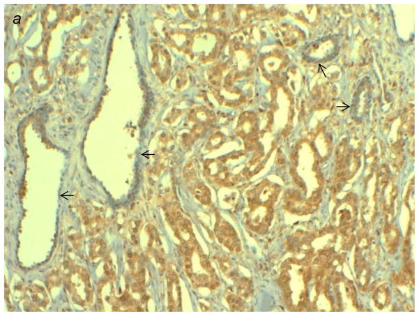
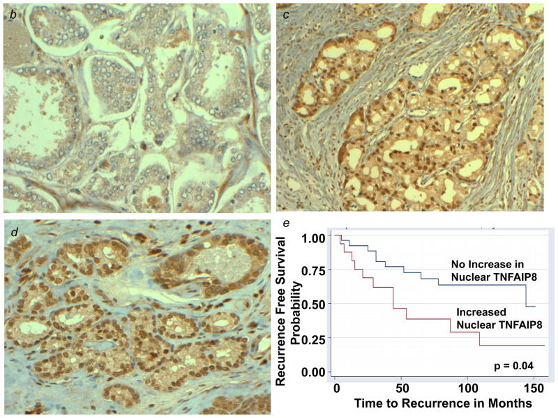
Figure 3.
(a) Immunostaining patterns of TNFAIP8 in high grade prostatic adenocarcinoma (PAC) versus adjacent benign glands (indicated by arrows). High grade PAC is showing intense cytoplasmic staining, whereas adjacent benign prostate shows weak, focal staining of TNFAIP8. (b–d) Expression and localization of TNFAIP8 in representative clinical specimens of PACs. (b) Low grade PAC showing weak to moderate cytoplasmic TNFAIP8, (c) high grade PAC showing intense cytoplasmic and intense nuclear TNFAIP8, and (d) high grade PAC showing intense nuclear TNFAIP8. (e) Kaplan-Meier survival curves estimates by nuclear TNFAIP8 immunoreactivity groups in prostatic adenocarcinoma patients.


