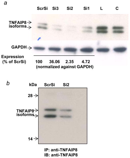
Figure 4.
(a) Verification of the siRNA silencing of TNFAIP8 in PC-3 cells by Western blotting. PC-3 cells were treated with 100 nM of the stealth TNFAIP8 siRNA (Si1, Si2, or Si3) or a scrambled stealth siRNA (ScrSi) for 4–6 hr, followed by incubation in complete IMEM for 72 hr as explained in Materials and Methods. The whole cell lysates were sequentially immunoblotted with anti-TNFAIP8 antibody (1:1000 dilution) and anti-GAPDH antibody. Data were quantified using NIH ImageJ software (version 1.45). C, untreated control; L, lipofectin-treated control. (b) Analysis of TNFAIP8 expression in TNFAIP8 immune-complexes from PC-3 cells treated with TNFAIP8 siRNA (Si2) versus Scrambled siRNA (ScrSi). PC-3 cells were treated with Si2 siRNA or Scr siRNA (ScrSi) as above and whole cell lysates (1.0 mg protein) were immunoprecipitated (IP) with anti-TNFAIP8, followed by immunoblotting (IB) with anti-TNFAIP8 antibody (1:1000 dilution).

