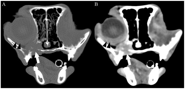
Figure 4.

Transverse 2mm collimated CT images of the orbits obtained 10 weeks after initial diagnosis and 4 weeks after exenteration of the left orbit of the cat shown in figure 1. Images are displayed with the left of the cat to the right of the image. (A) Pre-contrast image at the level of the right globe reconstructed with a bone filter and displayed with window width = 1500 HU and window level = 300 HU. (B) Post-contrast image reconstructed with a detail filter and displayed with a window width = 300 HU and window level = 100 HU. Note the contrast-enhancing soft tissue mass ventral to the right globe (white arrow) associated with osteolysis of the ventral and medial aspects of the orbit, palatine bone, and alveolar bone of the maxilla. The mass was contiguous with the episcleral and retrobulbar tissues caudally. The sclera, episclera, and retrobulbar tissues were thickened and markedly contrast enhancing (black arrowheads). The left eye was absent and replaced by a central non-contrast enhancing cystic structure (not shown) and thickened, contrast-enhancing peripheral soft tissues. HU, Hounsfield units.
