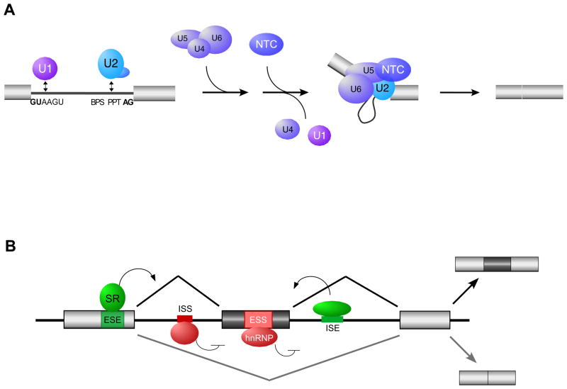
Fig. 2. General mechanism of splicing and its regulation.
(A) Spliceosome assembly. The 5′ end of introns are defined by the 5′ splice site (GUAAGU), and the 3′ end of introns are defined by the branch point sequence (BPS), polypirimidine tract (PPT), and the 3′ splice site AG dinucleotides. The U1, 2, 4, 5 and 6 represent the snRNPs which assemble with substrate and each other as shown. The NTC is an additional snRNA-free spliceosomal subunit. For more details see Motta-Mena et al. (58). (B) Regulation of alternative splicing. Enhancer auxiliary elements are denoted in green for exonic (ESE) or intronic (ISE) splicing enhancers. Silencer auxiliary elements are denoted in red for exonic (ESS) or intronic (ISS) splicing silencers. The activities of these auxiliary elements are often mediated through binding of SR and hnRNPs, two common families of proteins described in the text, among other RNA binding proteins.

