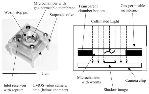
Fig. 12.

Photograph and schematic diagram of the lensless nematode shadow imager. Specimens are swimming within a 500 μm high chamber and are illuminated with an LED. The shadows of the objects are recorded using a CMOS image sensor attached to the bottom of the chamber. Reprinted from [64] with permission from Elsevier copyright (2005).
