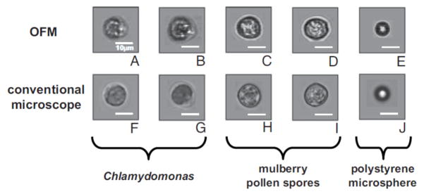
Fig. 16.

Cell and microsphere images captured by the electrokinetically driven OFM (A–E), and the corresponding microscope comparisons taken by a light transmission microscope with a 20× objective (F–J). Objects are Chlamydomonas (A,B and F,G), mulberry pollen spores (C,D, and H, I), and 10-μm polystyrene microspheres (E and J). Scale bars are 10 μm long. [71] Copyright (2008) National Academy of Sciences, USA.
