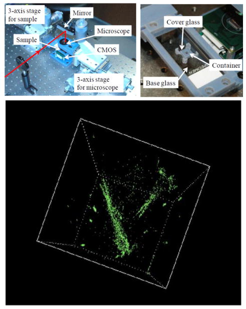
Fig. 17.
Photographs of the holographic microscopy system (top left) and the fluidic chamber in which water cyclopses are swimming (top right). A 3D image of the compressive holographic reconstruction of water cyclopses (bottom). The spatial resolution of this system is approximately 2.2 μm (lateral) together with an axial resolution of ~59 μm. Reprinted from [85] with permission from OSA.

