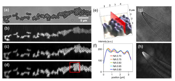
Fig. 3.

(a) Microscope image (0.75 NA) of 816nm PMMA beads. (b) DIHM image reconstructions of the same area using a highly coherent laser and (c) a partially coherent laser. (d) DIHM image reconstruction with an additional numerical correction of the glass sample carrier. (e) 3D view of the red framed image section in (d). (f) Sectional view for different NAs, as indicated in (e). (g) Optical microscope image (NA 0.75) of Pleurosigma angulatum and (h) the DIHM image reconstruction of the same object. Reprinted from [33] with permission from OSA.
