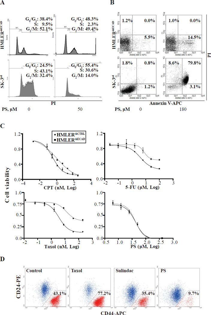
Fig. 1. PS effectively and selectively eliminates breast CSCs in vitro.
(A) The indicated cell lines were treated with 50 µM PS for 24 h, stained with propidium iodide and analyzed by flow cytometry for cell cycle phase distribution as in Methods. The experiment was repeated 3 times and representative results are shown. PS significantly reduced the percentage of cells in S phase in HMLERshECAD and SK-3rd (P < 0.05 compared with DMSO control). The percentage of cells in each phase is shown. (B) Cells were treated with 180 µM PS for 24 h, stained with propidium iodide and Annexin V-APC and analyzed by flow cytometry, as in Methods. Annexin V(+)/PI(-) = apoptosis; Annexin V(+)/PI(+) = end-stage apoptosis (secondary necrosis). The experiment was repeated 3 times and representative results are shown. PS significantly increased the percentage of dead HMLERshECAD and SK-3rd cells (P < 0.01 compared with DMSO control). (C) Inhibition curves and IC50s of each compound for the indicated cell lines treated with PS for 48 h Cell viability was determined by MTT assay as in Methods. (D) Sorted HMLER CD44high/CD24low and CD44low/CD24high cells were mixed at a ratio about 1:1 and plated in 6-well plates. After incubation with DMSO, PS (30 µM), Taxol (10 nM) or sulindac (700 µM) respectively for 7 days as in Methods, cells were stained for CD24/CD44 and analyzed by flow cytometry.

