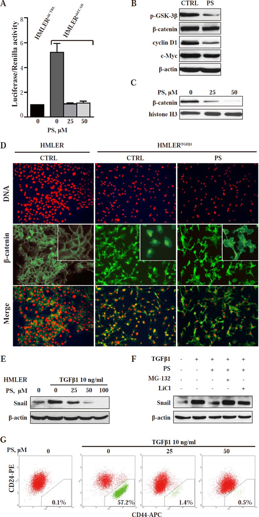
Fig.3. PS targets the Wnt/β-catenin pathway and reduces Snail expression blocking EMT and preventing the generation of CSCs.
(A) TCF/LEF transcriptional activity was tested by TOPFlash (TCF-LEF reporter) and FOPFlash (mutated binding sites). β-catenin/TCF-LEF activity is shown as TOP over FOP firefly luciferase activity, normalized to internal control Renilla luciferase activity. ***, P < 0.001 compared with control. (B) Immunoblot of the indicated proteins in HMLERshECAD cells after 36-h treatment with 30 µM PS. Loading control: β-actin. (C) Nuclear β- catenin levels in HMLERshECAD cells treated with PS determined by immunoblotting. Loading control: histone H3. (D) Immunofluorescence images of HMLER and HMLERTGFβ1 cells treated as shown for 24 h. Upper row: staining with PI visualizes the nuclei. Middle row: Staining with an 23 anti-β-catenin mAb. In HMLER cells, β-catenin is mainly in the cell membrane. In vehicle treated HMLERTGFβ1 cells, β-catenin is mainly nuclear/cytoplasmic, shifting predominantly to the cell membrane after PS treatment. Lower row: The merged images of the two rows above. Magnification: 20x. (E) HMLER cells were treated for 24 h with various concentrations of PS and with or without TGFβ1, the EMT-inducing agent. Protein levels of Snail were determined by immunoblotting. Loading control: β-actin. (F) HMLER cells were treated with the indicated compounds for 8 h. MG-132 or LiCl was added 10 min before other treatments started, as in Methods. (G) HMLER cells were incubated with TGFβ1 and the ind icated concentrations of PS for 14 days. CD24/CD44 expression profiles were then analyzed by flow cytometry as in Methods.

