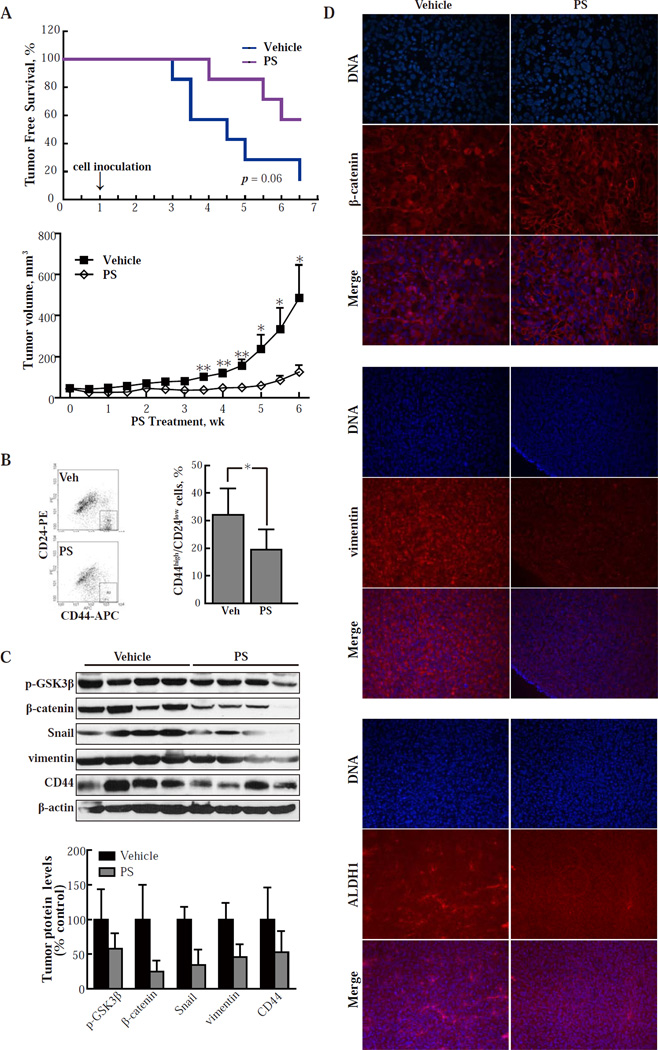
Fig. 4. PS inhibits the tumorigenicity of CSCs in nude mice.
(A) Upper panel: SK-3rd cells (2,000 per animal) were inoculated into immunodeficient mice. Treatment with vehicle or PS 100 mg/kg po daily 5x/wk started 1 wk prior to inoculation (prevention protocol). PS increased tumor-free survival by 50% (p=0.06; n=7/group). Lower panel: immunodeficient mice were inoculated with HMLERTGFβ1 cells (50,000/mouse) and treatment with vehicle or PS 150 mg/kg po daily 5x/wk started when the tumor reached the indicated size (treatment protocol). Values are mean±SEM; n=10/group. *, P < 0.05; **, P < 0.01 compared to control. (B) Single cell suspensions from HMLERTGFβ1 xenografts obtained by collagenase treatment as in Methods were stained for CD24/CD44 and analyzed by flow cytometry. In each group, 4 xenografts were studied and representative results are shown on the left. *, P < 0.05. (C) Protein levels in HMLERTGFβ1 xenografts were determined by immunoblotting and their band intensity (normalized to β-actin) is depicted in the graph. Values are mean±SD. *, P < 0.05; **, P < 0.01 compared to control. (D) Immunofluorescence images of tissue sections from HMLERTGFβ1 xenografts from the study depicted in A, lower panel (treatment protocol). Upper panel: staining with DAPI visualizes the nuclei and staining with an anti-β-catenin mAb reveals its distribution in the cells. The merged images of the two rows above are also shown. Analogous images from tissue sections stained for vimentin (middle panel) and ALDH1 (lower panel). Treatment with PS caused β-catenin to relocalize to the cell membrane. It also suppressed the expression of both vimentin and ALDH1, indicating suppression of EMT and near elimination of CSCs, respectively. Magnification: 20x.

