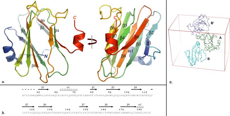
Fig. 1.
a. Two orthogonal views of the PYMOL 41-generated cartoon representation of the RetSperi structure. All secondary structure elements are labeled. b. Alignment of the secondary structure elements identified from the crystal structure with the primary structure RetSperi. This figure was generated with ESPript 42. c. The asymmetric unit of the crystal consists of molecules A and B. The contacts between A and B closely resemble the packing contacts between A and the symmetry-related B’ molecule, suggesting that the dimer in the asymmetric unit arose from crystal packing contacts.

