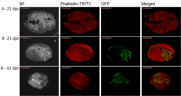
Figure 8.

Histochemical staining of HCT-116 tumor sections. Microscopic images of histological sections of representative HCT-116 tumors from an untreated mous at 21 days post-injection [A] and GLV-1h68-treated mice [B] at 21 and 42 days post-injection [dpi] are shown. Images were obtained under bright field (BF) or fluorescence microscopy (Phalloidin-TRITC, GFP). Digital images were processed with GIMP2 (Freeware) and merged to yield pseudocolored images (merged). Whole tumor cross-sections (thickness = 100 μm) were labeled with Phalloidin-TRITC to detect de novo synthesis of actin as an indicator for live cells. GFP fluorescence indicates GLV-1h68 replication in the tumor.
