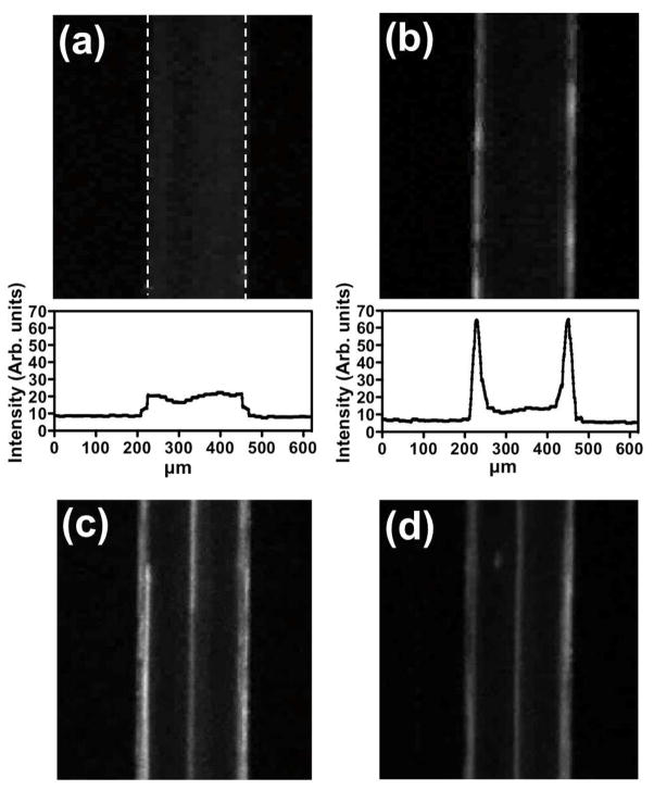
Figure 2.
ECμPs function as negative contrast particles and can be separated from positive contrast particles using an acoustic sample preparation chip. These images were captured via an epifluorescence microscope with a 2.5x objective. Each sample was flowing at 45 μL/min. Fluorescence microscopy images along with histograms of average intensity profiles for Nile Red (NR) stained ECμPs with the field (a) off and then (b) on. (c) NR-ECμPs separated from NR-PS particles with the field on. (d) NR-ECμPs separated from NR-blood cells with the field on. Note: white dashed lines in (a) indicate micro-channel borders.

