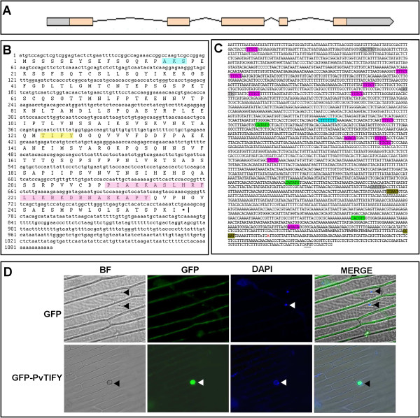
Figure 2.
Structural organization and cellular localization of PvTIFY10C. (A) PvTIFY10C gene structure. Exon regions are indicated with salmon-colored boxes, introns with black lines, and 5′ and 3′ UTRs with gray boxes. (B) Deduced amino acid sequence of PvTIFY10C. A predicted sumoylation site is shaded in blue. The TIFY domain is shaded in yellow. The Jas motive is shaded in pink. (C) The promoter region includes important regulatory cis-elements: CCAAT motifs (brown) at −15 and −713, G-boxes (CATATG; green) at −267, -911 and −1923, E-boxes (CANNTG; gray) at −798, 2474 and 3189, HUD (Hormone Up at Dawn; pink) elements at −41, -1304, -1563, -1642, -1655, -1947, -2785, -2788, -2791 and −3240, and a JA-responsive element (CTTTTNTC) at −1973. (D) PvTIFY10C is located in the nucleus. Onion epidermal cells were transiently transformed with a 35S:PvTIFY-GFP (GFP-TIFY) construct or with an empty vector (GFP). Epifluorescence (GFP, DAPI and MERGE) and bright-field (BF) images were captured of onion epidermal cells. Arrowheads indicate nuclei.

