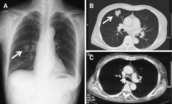
Figure 2.

Chest radiograph and computed tomography scan at diagnosis. (A) Chest radiograph showing a mass shadow in the right middle zone (arrow). (B, C) Selected sections of a conventional computed tomography scan of the chest showing a 30-mm solitary mass in S5 of the right lung (arrow), and mediastinal lymphadenopathy (arrowhead).
