Figure 16.

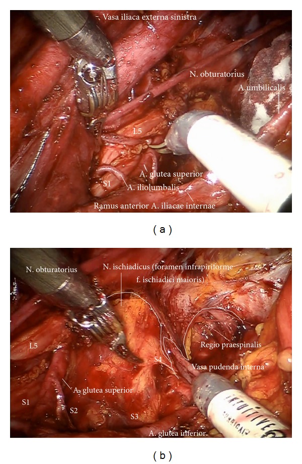
(a) Exposition of the superior gluteal artery and lumbosacral nerve roots (pv). (b) Visualization of the N. ischiadicus and the Vasa pudenda (upper paravisceral basin).

(a) Exposition of the superior gluteal artery and lumbosacral nerve roots (pv). (b) Visualization of the N. ischiadicus and the Vasa pudenda (upper paravisceral basin).