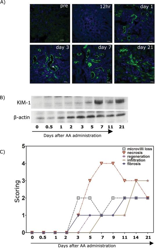
FIG. 4.

KIM-1 expression and histology in the mouse kidney after AA 5mg/kg administration. (A) Immunofluorescence staining of frozen kidney sections from AA-treated mice. Tissues were stained with KIM-1 antibody (R&D systems) as described in the Materials and Methods section. Tissue KIM-1 was clearly detected in 24h after AA (5mg/kg body weight) administration. (B) Western blot of the whole tissue kidney lysates collected from AA-treated (5mg/kg body weight) mouse. No KIM-1 was detected on day 0, but the protein levels became detectable by day 1 (24h) after AA administration. (C) Histology scoring of kidneys from AA-treated mice. Sections were evaluated for microvilli loss, necrosis, regeneration, inflammatory cell infiltration, and fibrosis on kidney tissues collected on day 0, 1, 2, 3, 5, 7, 11, and 21.
