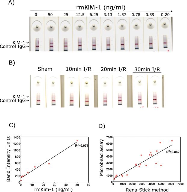
FIG. 8.
Detection of KIM-1 using mouse Rena-Stick in renal I/R model. KIM-1 in standard solutions or urine samples is measured by mixing 60 μl of sample with 60 μl of TRIS-buffered solution and adding the mix to the port in Rena-Stick. Results were read in 15min. (A) A concentration-dependent decrease in the band intensity was observed with a decrease in recombinant mouse KIM-1 protein standard concentration. (B) A visual increase in KIM-1 band intensity with an increase in bilateral ischemia time in mice subjected to renal I/R, whereas no KIM-1 positive band was observed when sham-operated mouse urine was analyzed. (C) The band intensity was quantified using a hand-held lateral-flow reader, and a standard curve was drawn by plotting the recombinant mouse KIM-1 concentration on x-axis and band intensity units on y-axis. (D) A positive correlation between KIM-1 values obtained by quantification of band intensity using the lateral-flow reader and values obtained by microbead-based assay in urines collected from bilateral I/R mice.

